
 Luiz Fernando Caserta Tencatt1
  , Alexandre Pucci Hercos2, Vandergleison de Carvalho3, Sérgio A. Santos4 and Marcelo R. Britto3
PDF: Download Here | Cite this article
Section Editor: William Crampton
Editor-in-chief: José Birindelli
Abstract
Uma nova espécie de Corydoras é descrita da bacia do rio Tefé, um tributário do rio Solimões, estado do Amazonas, Brasil. A nova espécie pode ser distinguida de suas congêneres por apresentar as seguintes características: uma mancha marrom-escura ou preta conspícua cruzando transversalmente a órbita, formando uma mancha semelhante a uma máscara; uma grande mancha marrom-escura ou preta transversalmente alongada que se estende da porção anterior da base da nadadeira dorsal até a margem ventral dos flancos, e ausência de um padrão de coloração distinto ao longo da linha mediana do flanco. Considerando que a nova espécie apresenta um padrão de coloração semelhante ao de Corydoras pastazensis, a identidade desta espécie também foi analisada neste estudo. O exame da maioria dos espécimes-tipo, incluindo holótipos, permitiu a clara delimitação de C. pastazensis, bem como a confirmação da sinonímia previamente proposta com C. pastazensis orcesi. Além disso, a análise da morfologia da boca e dos barbilhões em Corydoradinae indicou uma nova hipótese de homologia dos barbilhões, bem como a proposição de cinco novos caracteres relacionados a essas estruturas.
Palavras-chave: Barbilhão maxilar, Corydoras pastazensis, Osteologia, Rio Solimões, Taxonomia.
Introduction
The Callichthyidae armored catfishes can be promptly recognized among the Siluriformes by having two longitudinal series of dermal plates on the flanks (Reis, 1998, 2003). Representatives of this family can be found in most of the Neotropical region, occurring from rivers on the Pacific slope of Panama and extending to the La Plata basin in Argentina (Reis, 1998). Currently, the family harbors around 230 valid species distributed in two subfamilies, Callichthyinae and Corydoradinae, with five and seven valid genera, respectively (Dias et al., 2025; Fricke et al., 2025). Corydoradinae, which harbors approximately 91% of the family’s diversity, is composed of small-sized species with short maxillary barbel (not reaching the pectoral-fin origin), presenting wide variety of shapes and colors, which makes many of its representatives highly appreciated in aquarium trade (Reis, 1998, 2003; Tencatt, 2022).
Corydoras sensu Dias et al. (2025) is currently the third species-rich genus within Corydoradinae, with more than 30 valid species, behind Hoplisoma Swainson, 1838 and Brochis Cope, 1871, the first and second most speciose genera of the subfamily, respectively. The species of Corydoras Lacepède, 1803 can be distinguished from the remaining Corydoradinae genera by having the following features: (I) branch of the temporal sensory canal at sphenotic, which gives rise to the supraorbital canal, with two pores, (II) upper tooth plate of branchial arch with three or four series of teeth, (III) area at the corner of the mouth, ventral to the maxillary barbel, with a roughly triangular fleshy flap, which can be variably elongated, similar to a barbel, (IV) mesethmoid long, equal to or longer than entire length of frontal, (V) posterior margin of the dorsal-fin spine with antrorse serrations; (VI) posterior margin of the pectoral-fin spine with conical serrations, mostly strongly developed and retrorse, variably with some serrations perpendicularly directed or directed towards tip of spine, proximal and/or distal edge of spine variably with less-developed serrations, and (VII) posterior laminar expansion of infraorbital 2 contacting pterotic-extrascapular (Dias et al., 2025; Tencatt et al., 2025a).
The number of putatively undescribed species of Corydoradinae in the aquarium trade is remarkable, especially from the rio Amazonas basin (see Tencatt et al., 2021, 2022a, 2023a,b, 2024a,b, 2025a). Currently, there are 48 putatively undescribed species coded in the aquarium. Among the coded species stands Corydoras sp. CW128, reported from the Quebrada Saragossa close to the city of Nauta in Peru, and CW207, reported from the rio Içá in the region of the border between Brazil and Colombia, both tributaries of the rio Amazonas. These species possess a peculiar color pattern, composed of a dark mask-like blotch transversally crossing the orbit and a large, transversally elongated blotch extending from the anterior portion of the dorsal-fin base toward the ventral margin of the trunk, with remaining portions of flank covered only with small dark brown or black blotches. Such color pattern is reminiscent to the one displayed by C. pastazensis Weitzman, 1963, a species described from the rio Pastaza basin in Ecuador. Currently, the delimitation of C. pastazensis is unclear, mostly due to the doubtful taxonomic status of C. orcesi Weitzman & Nijssen, 1970, originally described as its subspecies. Corydoras pastazensis orcesi, which, according to Weitzman, Nijssen (1970), differs from C. pastazensis pastazensis by having some differences in color pattern, was described from the same region in Ecuador as C. pastazensis, but from a tributary of the rio Tigre.
Diverging from their broader taxonomic review of Corydoras (Nijssen, Isbrücker, 1980a), which considered C. pastazensis orcesi as a valid subspecies, Nijssen, Isbrücker (1986) placed C. pastazensis orcesi in the synonymy of C. pastazensis in their taxonomic review of the Corydoras species from Peru and Ecuador. The authors discussed differences in color pattern between C. pastazensis pastazensis and C. pastazensis orcesi, and of similar specimens from the drainages of the rivers Napo in Ecuador and Ucayali in Peru. No explanation or arguments were given for the synonymy. Although no review work has proposed the revalidation of C. pastazensis orcesi, at the subspecies or species level, the use of the name C. orcesi was arbitrarily brought up in some publications (e.g., Isbrücker, 2001; Alexandrou et al., 2011; Lima, Sazima, 2017).
The analysis of the holotype (USNM 177216) and three paratypes (USNM 164464) of C. pastazensis, as well as of the holotype (USNM 204358) and seven paratypes of C. pastazensis orcesi (USNM 203827, 203828) allowed us to confirm the synonymy proposed by Nijssen, Isbrücker (1986). Additionally, the analysis of specimens similar to Corydoras sp. CW128 and CW207 from the rio Tefé, Amazonas State, revealed them as representatives of an undescribed species, which is formally described herein. Nonetheless, the new species has some differences in color pattern when compared to Corydoras sp. CW128 and CW207, suggesting that they may represent variable forms of C. pastazensis or even undescribed species. Comments on the identity of both coded species are provided herein.
Material and methods
The classification of Corydoradinae and generic placement of the new species follows Dias et al. (2025), according to the reasoning provided by Tencatt et al. (2025b). Measurements were obtained using digital calipers to the nearest tenth of millimeter. Morphometric and meristic data were taken following Tencatt et al. (2022b) and Reis (1997), respectively. Morphometrics are reported as percent of standard length (SL) or head length (HL). Terminology of barbels follows Britto, Lima (2003), but with the homology proposed herein. Orientation of the serrations on the posterior margins of the dorsal- and pectoral-fin spines followed terminology according to Ballen, de Pinna (2021). For the osteological analysis, some specimens were cleared-and-stained (c&s) according to the protocol of Taylor, Van Dyke (1985). Due to fading of bone staining, some paratypes were re-stained in a solution of 75% ethyl alcohol and alizarin red S (Springer, Johnson, 2000) after dissection. Osteological terminology was based on Reis (1998), except for the use of parieto-supraoccipital instead of supraoccipital (Arratia, Gayet, 1995), pterotic-extrascapular instead of pterotic-supracleithrum (Slobodian, Pastana, 2018), and scapulocoracoid instead of coracoid (Lundberg, 1970). Nomenclature of the latero-sensory canals and preopercular pores are according to Schaefer, Aquino (2000) and Schaefer (1988), respectively. The supra-preopercle sensu Huysentruyt, Adriaens (2005) was treated here as a part of the hyomandibula according to Vera-Alcaraz (2013). To determine the development degree of the anterior laminar expansion of infraorbital 1 in relation to the nasal capsule, the specimen was positioned to maintain the largest diameter of the nasal capsule horizontally. The width of frontal bone was obtained at the same point as the least interorbital width, following Tencatt et al. (2022b). Vertebral counts include only free centra (i.e., post-Weberian complex), with the compound caudal centrum (preural 1+ ural 1) counted as a single element. The relative size of dorsal-, pectoral-, pelvic- and anal-fin rays are analyzed qualitatively, comparing each ray in erected position (fin fully abducted), with the largest fin element as the main reference. The last two dorsal-fin rays were counted as distinct elements. Pharyngeal teeth were counted on both sides of the branchial arches. In the photos illustrating c&s specimens, the apparent size of some structures may eventually be altered by the perspective from which the photo was taken (e.g., it was not possible to perfectly align the mesethmoid in the horizontal plane in all photographed specimens, especially in dorsal view).
In the description, numbers in parentheses represent the total number of specimens with those counts. Numbers with an asterisk refer to the counts of the holotype. In the color pattern descriptions, the longitudinal markings on the body are referred to as stripes, whereas the transversal markings on the head and trunk are referred to as bars; the transversal markings on fins are referred to as bands. Institutional abbreviations follow Sabaj (2025). The photograph of the specimen NRM 28578 mentioned in the Remarks section can be visualized in https://samlingar.nrm.se/record/pi_28578. Part of the photographs of the specimens LBP 2835, 57.8 mm SL, and LBP 2836, 41.7 mm SL, as well as the LBP collection database were directly obtained from the curator, Claudio Oliveira. Political geography data of the type material is presented in the following order: country, state and municipality. The conservation status of the new species was suggested using the categories and criteria of the International Union for Conservation of Nature guidelines (IUCN Standards and Petitions Subcommittee, 2024). General morphological information on Callichthyinae were retrieved from Reis (1997, 1998).
Results
Corydoras isaacisbrueckeri, new species
urn:lsid:zoobank.org:act:35B330A8-D046-4C2A-8601-2223C9A6FBFB
(Fig. 1; Tab. 1)
Holotype. MNRJ 56008, 58.3 mm SL, Brazil, Amazonas, Tefé, igarapé da Associação dos Trabalhadores do Mamirauá (ATM), a tributary of the rio Tefé, rio Solimões basin, 03°27’13”S 64°40’22”W, 1 Nov 2024, D. Mendes, J. Oliveira & J. Oliveira.
Paratypes. All from Brazil, Amazonas, Tefé, rio Tefé basin, rio Solimões basin. CITL 1557, 4 of 6, 38.1–57.7 mm SL, 2 c&s of 6, 40.9–43.0 mm SL; INPA 62037, 4, 36.1–48.0 mm SL; MUBIO 722, 3, 41.6–49.0 mm SL; MZUSP 130976, 4, 41.4–54.6 mm SL; NUP 26023, 4, 42.3–47.7 mm SL, igarapé da Barreira, 04°17’52”S 65°12’22”W, 23 Jun 2024, A. P. Hercos and E. G. Silva. IDSM 4699, 2, 33.6–51.3 mm SL, igarapé do Pavão, 03°31’24”S 64°37’56”W, 7 Sep 2024, R. M. Holanda, D. Mendes & J. Oliveira. IDSM 4700, 1, 61.9 mm SL, collected with the holotype.

FIGURE 1| Corydoras isaacisbrueckeri, holotype, MNRJ 56008, 58.3 mm SL, Tefé, Amazonas, Brazil, igarapé da Associação dos Trabalhadores do Mamirauá (ATM), a tributary of the rio Tefé, rio Solimões basin.
Diagnosis. Corydoras isaacisbrueckeri can be distinguished from its congeners, except for C. amapaensis Nijssen, 1972, C. blochi Nijssen, 1971, C. caramater Tencatt, Couto, Santos & Sousa, 2024, C. cortesi Castro, 1987, C. desana Lima & Sazima, 2017, C. iiap Tencatt, Ruiz-Tafur & Chuctaya, 2024, C. pastazensis Weitzman, 1963, C. saramaccensis Nijssen, 1970, C. septentrionalis Gosline, 1940, C. serratus Sands, 1995, C. solox Nijssen & Isbrücker, 1983,and C. simulatus Weitzman & Nijssen, 1970, by having a conspicuous dark brown or black patch transversally crossing the orbit, forming a mask-like blotch (vs. mask-like blotch absent); it differs from C. amapaensis, C. blochi, C. caramater, C. cortesi, C. iiap, C. saramaccensis, C. septentrionalis, C. serratus, C. solox, and C. simulatus by having a large, transversally elongated dark brown or black patch extending from the anterior portion of the dorsal-fin base toward the ventral margin of the trunk (vs. dark patch on the anterior portion of the dorsal-fin base absent in C. amapaensis, C. caramater, C. iiap, and C. solox; dark patch on the anterior portion of the dorsal-fin base roughly rounded, not extended ventrally in C. blochi, C. cortesi, C. saramaccensis, C. septentrionalis, and C. serratus; dark patch on the anterior portion of the dorsal-fin, when present, roughly rounded, not extended ventrally in C. simulatus); it differs from C. desana and C. pastazensis, plus C. cortesi, C. septentrionalis, and C. simulatus by the absence of a distinct color pattern along the midline of the flank (vs. midline of the flank with at least two moderate- to large-sized, conspicuous dark brown or black longitudinally aligned blotches in C. desana, C. pastazensis, and C. septentrionalis; midline of the flank typically with two moderate- to large-sized, conspicuous dark brown or black longitudinally aligned blotches, some specimens variably with a single blotch in C. simulatus; midline of flank with a dark brown or black stripe in C. cortesi).
Description. Morphometric data in Tab. 1. Head laterally compressed with acutely convex dorsal profile, roughly triangular in dorsal view. Snout well developed, conical; conspicuously pointed in some specimens. Head profile slightly concave from tip of snout to anterior nares; nearly straight in some specimens; ascending nearly straight to slightly convex from that point to dorsal-fin origin; region of frontal fontanel slightly concave in some specimens. Profile nearly straight to slightly convex along dorsal-fin base. Postdorsal-fin body profile slightly concave to adipose-fin spine, slightly concave from this point to caudal-fin base; region between dorsal and preadipose platelets typically roughly straight. Ventral profile of body nearly straight or slightly convex from isthmus to pectoral girdle, and slightly convex from this point until pelvic girdle. Profile nearly straight to slightly convex from pelvic girdle to base of first anal-fin ray, ascending slightly concave until caudal-fin base. Body roughly elliptical in cross section at pectoral girdle, gradually becoming more compressed toward caudal fin. Highest body depth at vertical through anterior dorsal-fin origin.
TABLE 1 | Morphometric data of the holotype and 19 paratypes of Corydoras isaacisbrueckeri. SD = Standard deviation.
| Holotype | Low–High | Mean±SD |
Standard length (mm) | 58.3 | 36.1–58.3 | 47.2 |
Percent of standard length | |||
Depth of body | 34.8 | 34.3–40.3 | 37.1±1.6 |
Predorsal distance | 50.8 | 47.8–53.2 | 50.7±1.3 |
Prepelvic distance | 46.5 | 46.3–49.6 | 47.5±0.8 |
Preanal distance | 79.2 | 78.5–83.7 | 80.2±1.2 |
Preadipose distance | 82.5 | 81.4–84.9 | 83.0±1.0 |
Length of dorsal spine | 21.6 | 21.6–26.2 | 23.9±1.2 |
Length of pectoral spine | 22.6 | 22.4–26.3 | 24.4±1.2 |
Length of adipose-fin spine | 9.6 | 8.1–11.8 | 9.9±0.9 |
Depth of caudal peduncle | 13.2 | 13.2–15.7 | 14.7±0.8 |
Length of dorsal-fin base | 18.4 | 18.4–21.1 | 20.0±0.7 |
Dorsal to adipose distance | 18.0 | 15.3–19.1 | 17.0±1.0 |
Maximum cleithral width | 23.8 | 22.6–25.4 | 23.8±0.8 |
Length of maxillary barbel | 18.5 | 17.6–21.8 | 19.3±1.1 |
Head length | 43.7 | 41.9–45.8 | 44.1±1.1 |
Percent of head length | |||
Head depth | 73.3 | 73.3–83.4 | 78.5±2.3 |
Least interorbital distance | 21.2 | 19.1–23.3 | 21.3±0.9 |
Horizontal orbit diameter | 18.8 | 18.8–23.7 | 21.6±1.4 |
Snout length | 50.6 | 45.3–50.7 | 48.1±1.6 |
Least internarial distance | 10.6 | 10.1–13.0 | 11.1±0.8 |
Eye rounded, located dorsolaterally on head. Orbit delimited anteriorly by lateral ethmoid, anterodorsally by frontal, posterodorsally by sphenotic, posteroventrally by infraorbital 2, and anteroventrally by infraorbital 1 (Fig. 2). Anterior and posterior nares close to each other, separated only by flap of skin. Anterior naris tubular. Posterior naris close to anterodorsal margin of orbit, separated from it by distance similar to naris diameter. Mouth small, subterminal, width similar to bony orbit diameter; skin fold extending from region just dorsolateral to mouth towards anteroventral portion of preopercle, forming lateral fold at anteroventral portion of snout; posterior portion of lateral fold expanded laterally, forming ventrolateral groove; area at mouth corner, just ventral to anterior portion of lateral fold, with small, roughly triangular fleshy flap; posterolateral portion of mouth bulbous (i.e., maxillary bulb), with posteroventral margin slightly projected laterally; posterolateral margin of lower lip poorly developed, roughly straight to slightly rounded (Fig. 3). Outer mental barbel typically well developed, reaching to or slightly surpassing anteroventral limit of gill opening; moderately developed, nearly reaching anteroventral limit of gill opening in some specimens, apparently due to ongoing regeneration; dorsolateral skin fold from anterolateral portion of barbel to middorsal portion of maxillary bulb. Maxillary barbel typically similar in size to outer mental barbel, variably slightly shorter or longer than outer mental barbel; small, roughly triangular to rounded mesial skin fold from posteroventral portion of upper lip to anteroventral portion of barbel. Inner mental barbel fleshy, base of each counterpart slightly separated from each other (Fig. 3). Small rounded papillae covering entire surface of all barbels, upper and lower lips, snout and isthmus.
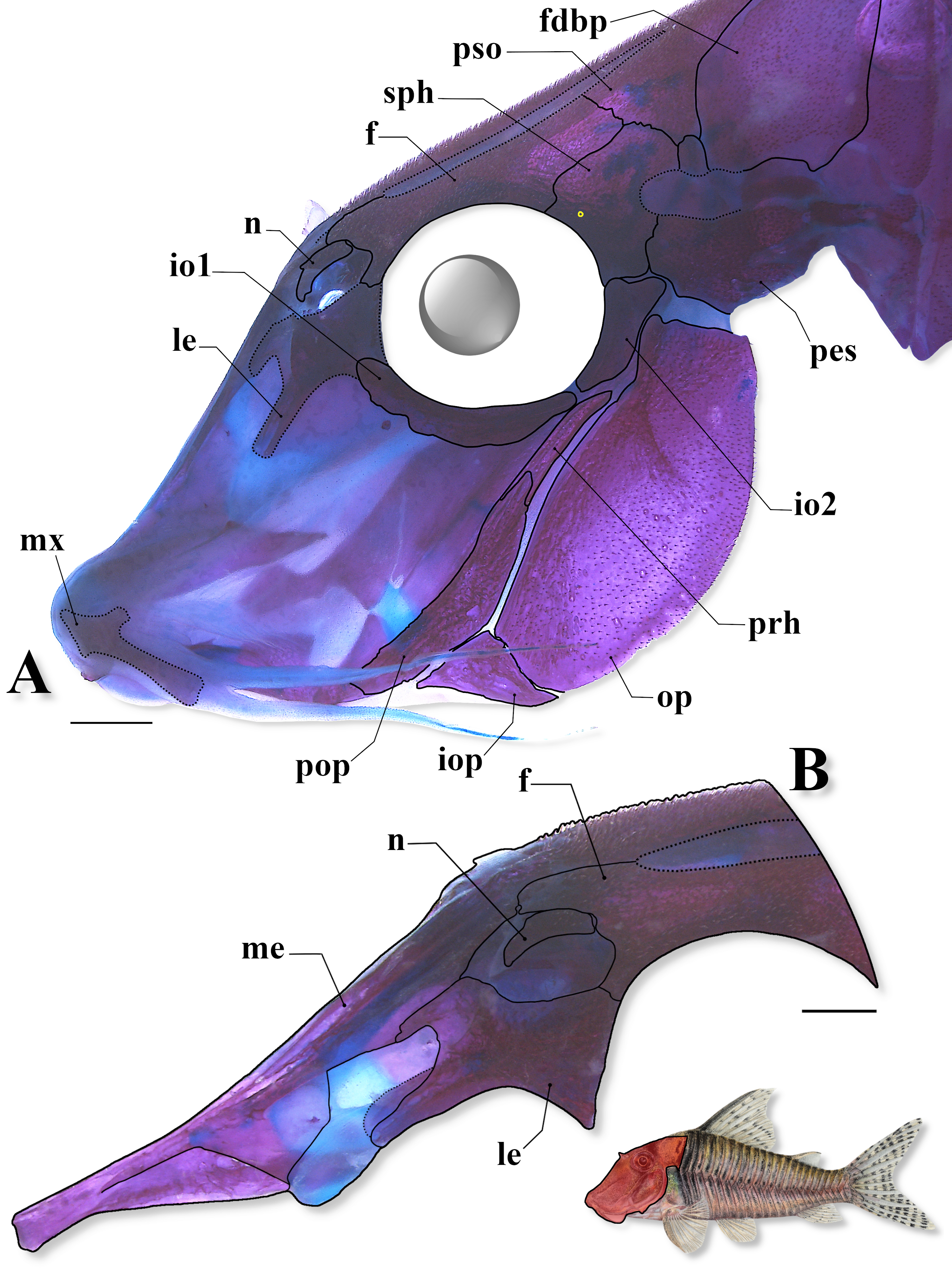
FIGURE 2| Head osteological pattern in a c&s paratype of Corydoras isaacisbrueckeri (CITL 1557, 43.0 mm SL), showing (A) general morphology in lateral view, and (B) the anterior portion of neurocranium in lateral view. Abbreviations: f: frontal, fdbp: first dorsolateral body plate, io1–2: infraorbital 1 and 2, iop: interopercle, le: lateral ethmoid, n: nasal, me: mesethmoid, mx: maxilla, op: opercle, pes: pterotic-extrascapular, pop: preopercle, prh: posterodorsal ridge of hyomandibula, pso: parieto-supraoccipital, sph: sphenotic. Additional pore of the temporal sensory canal at sphenotic outlined in yellow. Area where the illustrated bones are located in fish’s body marked in red in the miniature drawing of the new species. Scale bars = 1 mm.
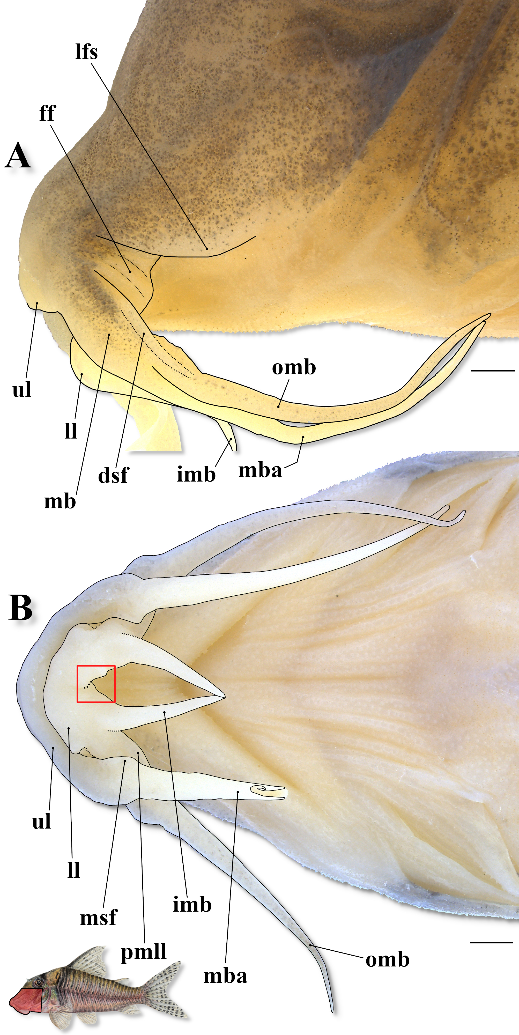
FIGURE 3| Detail of the snout and mouth in (A) a paratype of Corydoras isaacisbrueckeri (CITL 1557, 54.5 mm SL), and (B) ventral surface of head in a paratype of C. isaacisbrueckeri (MZUSP 130976, 47.7 mm SL), showing the mouth-related structures (outlined in black) of new species. Abbreviations: dsf: dorsolateral skin fold of the maxillary bulb, ff: fleshy flap, imb: inner mental barbel, lfs: lateral fold at anteroventral portion of snout, ll: lower lip, mb: maxillary bulb, mba: maxillary barbel, mfs: mesial skin fold of the maxillary barbel, omb: outer mental barbel, pmll: posterolateral margin of lower lip, ul: upper lip. Red square highlighting the region of the mesial notch of the lower lip. Area where the illustrated structures are located in fish’s body marked in red in the miniature drawing of the new species. Scale bars = 1 mm.
Mesethmoid long, its length larger than frontal length; anterior tip smoothly inclined upwards in lateral view, relatively short, slightly smaller than 50% of bone length; posterior margin relatively narrow, its width smaller than maximum width of posterior portion of bone; base relatively short, lateral posterodorsal expansion with relatively wide roughly trapezoid external projection, emerging relatively close to distal point of suture with frontal; external projection relatively short, its distal tip not reaching outermost margin of nasal bone; posterodorsal portion entirely covered by thick layer of skin; posterior ventrolateral expansion clearly visible in dorsal view, emerging on middle portion of mesethmoid, further anteriorly to external projection of lateral posterodorsal expansion (Fig. 4). Upper and lower jaws edentulous; premaxilla overall funnel-like shaped, with anteroventral surface roughly horizontally rectangular in frontal view; anteroventral margin irregular; posterodorsal portion with conspicuous pointed process, mesially set in frontal view (Fig. 5); maxilla elongated, relatively slender and roughly hatchet shaped in frontal view, its proximal half with roughly trapezoid laminar process on posterolateral portion (Fig. 5); dentary relatively slender, with roughly trapezoid expansion on its anteroventral portion, in ventrolateral perspective, perpendicularly directed or smoothly bent anteriorly; roughly triangular process on its posterodorsal portion, bent posteriorly; process strongly reduced, almost imperceptible on left side of specimen CITL 1557, 43.0 mm SL; angulo-articular relatively deep posteriorly, with roughly triangular dorsal laminar expansion, its posterodorsal margin typically irregular; dorsal laminar expansion bent posteriorly; posteroventral portion with roughly triangular process, in lateral view, bent posteriorly (Fig. 5). Palatine longitudinally elongated, slender, with roughly triangular dorsolateral longitudinal laminar expansion, gradually increasing in depth posteriorly, articulating with lateroventral expansion of posterior portion of mesethmoid; posterolateral process well developed, extending posteriorly in parallel to dorsal margin of anterior laminar expansion of metapterygoid (Fig. 5).
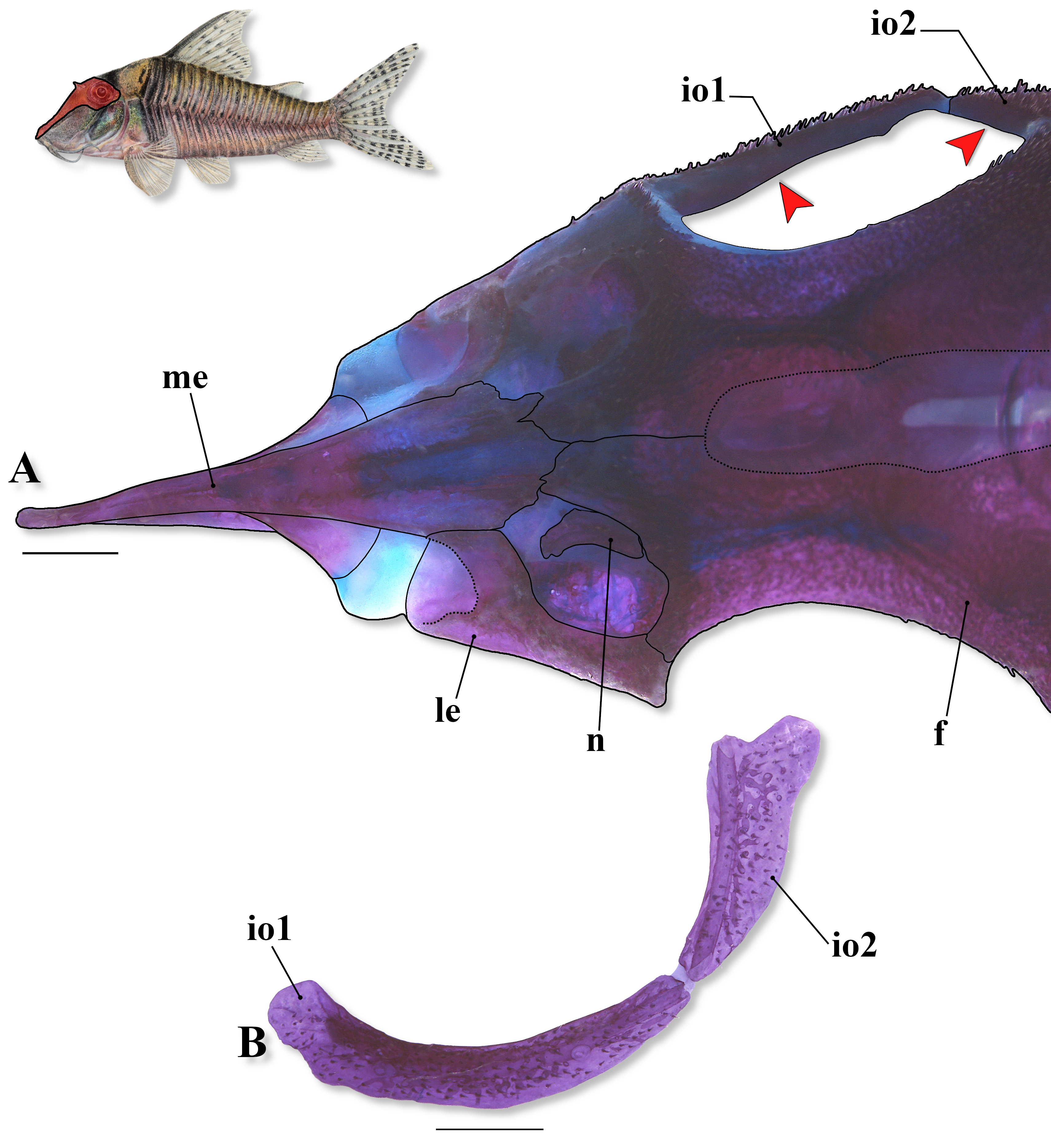
FIGURE 4| Cranial osteology in a c&s paratype of Corydoras isaacisbrueckeri (CITL 1557, 43.0 mm SL), showing (A) the anterior portion of neurocranium in dorsal view, and (B) the infraorbital series in lateral view. Abbreviations: f: frontal, io1–2: infraorbital 1 and 2, le: lateral ethmoid, me: mesethmoid, n: nasal. Red arrows indicate inner laminar expansion of both infraorbitals. Area where the illustrated bones are located in fish’s body marked in red in the miniature drawing of the new species. Scale bars = 1 mm.
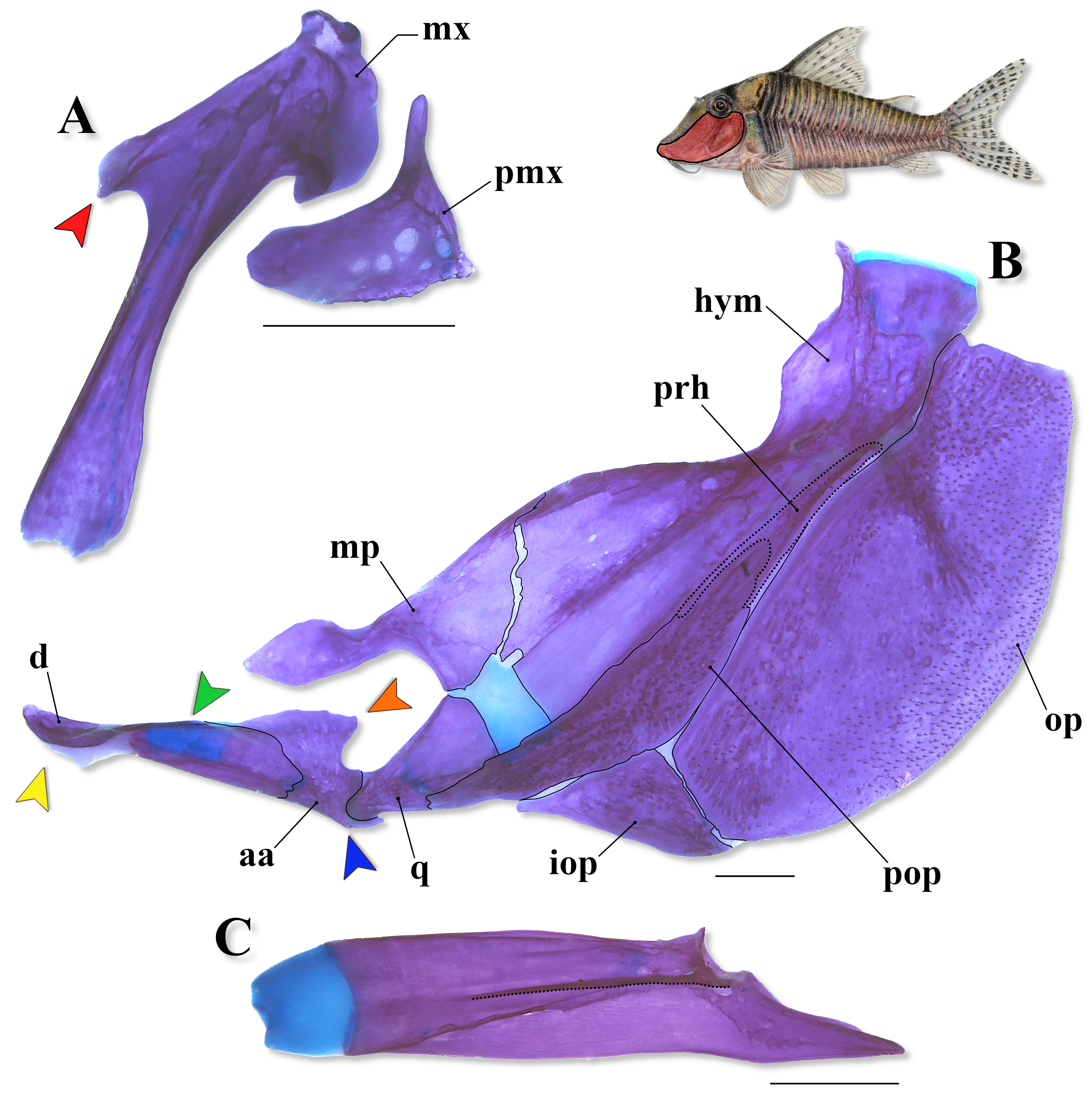
FIGURE 5| Cranial osteology in a c&s paratype of Corydoras isaacisbrueckeri (CITL 1557, 43.0 mm SL), showing (A) the upper jaw in frontal view (= dorsal view considering everted mouth), (B) the suspensorium plus operculum in lateral view, and (C) the palatine in dorsal view. Abbreviations: aa: angulo-articular, d: dentary, hym: hyomandibula, iop: interopercle, mp: metapterygoid, mx: maxilla, op: opercle, pmx: premaxilla, pop: preopercle, prh: posterodorsal ridge of hyomandibula, q: quadrate. Red arrow indicates laminar process on posterolateral portion of maxilla; yellow and green arrows indicate anteroventral expansion (covered by thick layer of skin; not visible) and posterodorsal process (strongly reduced) of dentary, respectively; orange and blue arrows indicate dorsal laminar expansion and posteroventral process of angulo-articular, respectively. Dotted line in (C) outlining dorsolateral longitudinal laminar expansion of palatine. Area where the illustrated bones are located in fish’s body marked in red in the miniature drawing of the new species. Scale bars = 1 mm.
Nasal capsule delimited posterodorsally and dorsally by frontal, anterodorsally by mesethmoid, and anteroventrally, ventrally and posteroventrally by lateral ethmoid (Fig. 2). Nasal slender, laterally curved, inner margin typically with poorly-developed laminar expansion, contacting only frontal; outer margin with strongly reduced laminar expansion (Figs. 2, 4). Lateral ethmoid overall compact, deep in lateral view, entirely covered by thick skin layer; posterior portion with relatively wide roughly triangular posteroventral expansion, and relatively short and slender roughly trapezoid posterodorsal expansion; distinct curved anteriorly anterodorsal expansion, its posterodorsal portion relatively distant from nasal, and anterodorsal margin contacting external projection of lateral posterodorsal expansion of mesethmoid; anteroventral expansion strongly well developed, conspicuously expanded anteroventrally, contacting ventrolateral expansion of posterior portion of mesethmoid by means of large cartilage block (Figs. 2, 4). Frontal elongated, extremely narrow, width clearly smaller than half of entire length, anterior projection partially covered by thick layer of skin; anterolateral process roughly trapezoid, slender in lateral view (Figs. 2, 4). Frontal fontanel large, slender, and somewhat ellipsoid; posterior tip extension clearly surpassing anterior margin of parieto-supraoccipital (Figs. 2, 4). Sphenotic somewhat trapezoid, contacting parieto-supraoccipital dorsally, pterotic-extrascapular posteriorly, second infraorbital posteroventrally and frontal anteriorly (Fig. 2). Pterotic-extrascapular roughly pipe-shaped, with posteriormost portion contacting first lateral-line ossicle, posteroventral margin contacting cleithrum, and anteroventral margin contacting opercle and infraorbital 2; posterior expansion almost entirely covering lateral opening of swimbladder capsule, leaving slender area on its dorsal margin covered only by thick layer of skin (Fig. 2). Parieto-supraoccipital wide, posterior process long, its length larger than half of parieto-supraoccipital length; contacting nuchal plate; region of contact between posterior process and nuchal plate covered by thick layer of skin.
Two laminar infraorbitals with minute odontodes. Infraorbital 1 large, ventral laminar expansion ranging from poorly to well developed; depth of ventral laminar expansion apparently related with specimen size, increasing in size during growth; anterior portion with laminar expansion ranging from poorly developed, slightly surpassing posterior margin of nasal capsule, to conspicuously well-developed, slightly surpassing anterior margin of nasal capsule; length of anterior laminar expansion apparently related with specimen size, increasing in size during growth; inner laminar expansion strongly reduced (Figs. 2, 4). Infraorbital 2 small, widened dorsally, with posterior laminar expansion ranging from moderately to well developed (Figs. 2, 4); posteroventral margin contacting posterodorsal ridge of hyomandibula, posterior margin contacting opercle, and posterodorsal edge contacting sphenotic and pterotic-extrascapular (Fig. 2); posterodorsal portion with roughly rounded expansion, and middle portion with nearly straight to smoothly rounded expansion (Figs. 2, 4); inner laminar expansion strongly reduced (Fig. 4). Posterodorsal ridge of hyomandibula close to its articulation with opercle slender, exposed, and bearing small odontodes (Figs. 2, 5). Dorsal ridge of hyomandibula between pterotic-extrascapular and opercle covered by thick layer of skin. Interopercle entirely or partially covered by thick layer of skin; posterior portion variably exposed and bearing odontodes; subtriangular, anterior projection moderately developed (Figs. 2, 5). Preopercle elongated, relatively slender; minute odontodes on external surface (Figs. 2, 5). Opercle dorsoventrally elongated, with width slightly smaller than half of its entire length; free margin convex, posterodorsal portion with smoothly concave area in some specimens, posteroventral margin variably irregular; without serrations and covered by small odontodes (Figs. 2, 5).
Four branchiostegal rays decreasing in size posteriorly. Hypobranchial 1 deep; hypobranchial 2 subtriangular, tip ossified and directed towards anterior portion, posterior margin cartilaginous; ossified portion moderately developed, its size similar to cartilaginous portion; paratype CITL 1557, 40.9 mm SL, with ossified portion of hypobranchial 2, except for its tip, covered by thin layer of cartilage. Five ceratobranchials with expansions increasing posteriorly; ceratobranchial 1 with reduced process on anterior margin of mesial portion; ceratobranchial 3 with continuous laminar expansion on postero-lateral margin; ceratobranchial 5 toothed on posterodorsal surface, with 27 to 29 (2) teeth aligned in one row. Four epibranchials with similar size; epibranchial 2 slightly larger than others, with small, roughly trapezoid process on laminar expansion of posterior margin, variably with notch just ventral to process; epibranchial 3 with roughly triangular uncinate process on laminar expansion of posterior margin; process variably bent mesially. Two wide pharyngobranchials (3 and 4); pharyngobranchial 3 with roughly trapezoid laminar expansion on posterior margin; laminar expansion variably notched. Upper tooth plate roughly oval, 50 to 54 (2) teeth aligned in three rows on posteroventral surface; rows closely aligned.
Lateral-line canal reaching cephalic laterosensory system through pterotic-extrascapular, branching twice before reaching sphenotic: pterotic branch, with single pore, preoperculomandibular branch conspicuously reduced, with single pore opening at postotic main canal; postotic main canal widens just posterior to pterotic branch. Sensory canal continuing through pterotic-extrascapular, reaching sphenotic as temporal canal, which splits into two branches: one branch giving rise to infraorbital canal, other branch connecting to frontal through supraorbital canal, with one and two pores, respectively. Supraorbital canal branched, running through nasal bone. Epiphyseal branch relatively long; pore opening close to frontal fontanel. Nasal canal typically with three openings, first on posterior edge, second on posterolateral portion, fused with first pore, and third on anterior edge. Infraorbital canal running through entire infraorbital 2, extending to infraorbital 1 and typically opening into two pores. Preoperculomandibular branch giving rise to preoperculo-mandibular canal, which runs through almost entire preopercle with three openings, leading to pores 3, 4, and 5, respectively.
Dorsal fin subtriangular, located just posterior to second or third dorsolateral body plate. Dorsal-fin rays II,8*(20), with first branched ray as longest fin element; branched rays typically decreasing in size posteriorly, with first and second, and variably third, branched rays slightly longer than ossified portion of dorsal-fin spine, remaining rays with similar size or shorter than spine; second branched ray similar in size to ossified portion of spine in some specimens; first and second branched rays clearly more elongated than spine in some specimens; posterior margin of dorsal-fin spine with six strongly reduced to poorly-developed serrations in both c&s paratypes (CITL 1557); most serrations antrorse, restricted to distal half of spine; some serrations variably perpendicularly directed (relative to main axis of spine); small odontodes on anterior and lateral surfaces of spine (Fig. 6). Nuchal plate well developed, overall size similar to posterior process of parieto-supraoccipital; almost entirely exposed, with minute odontodes. Spinelet short; spine typically moderately developed, with adpressed distal tip reaching to or slightly surpassing posterior origin of dorsal-fin base. Pectoral fin roughly triangular, its origin just posterior to gill opening. Pectoral-fin I,10(8), I,10,i(4), I,11*(8), with first branched ray typically as longest fin element, second branched ray variably similar in size to first branched ray; branched rays decreasing in size posteriorly, with first, second and third branched rays slightly typically longer than ossified portion of pectoral-fin spine, remaining rays with similar size or shorter than spine; second and/or third branched rays variably similar in size to spine (apparently due to ongoing regeneration in some specimens); posterior margin of pectoral spine with 13 to 24 conical serrations along almost its entire length, absent around origin of spine; most serrations well developed and retrorse; serrations close to origin of spine conspicuously less developed; some serrations, especially on proximal portion of spine, variably perpendicularly directed (relative to main axis of spine) or antrorse; bifid serrations variably present; small odontodes on anterior, dorsal and ventral surfaces of spine (Fig. 6). Anteroventral portion of cleithrum and anterolateral portion of scapulocoracoid exposed; posterolateral portion of scapulocoracoid moderately developed, exposed, with anterior portion slightly to moderately expanded anteriorly, not in contact with anteroventral portion of cleithrum; larger specimens (with more than 50.0 mm SL) with posterolateral portion of scapulocoracoid moderately expanded mesially; exposed areas bearing small odontodes. Opening of axillary gland sensu Kiehl et al. (2006) located just posterior to pectoral-fin spine base; opening apparently reduced to narrow slit in some specimens.
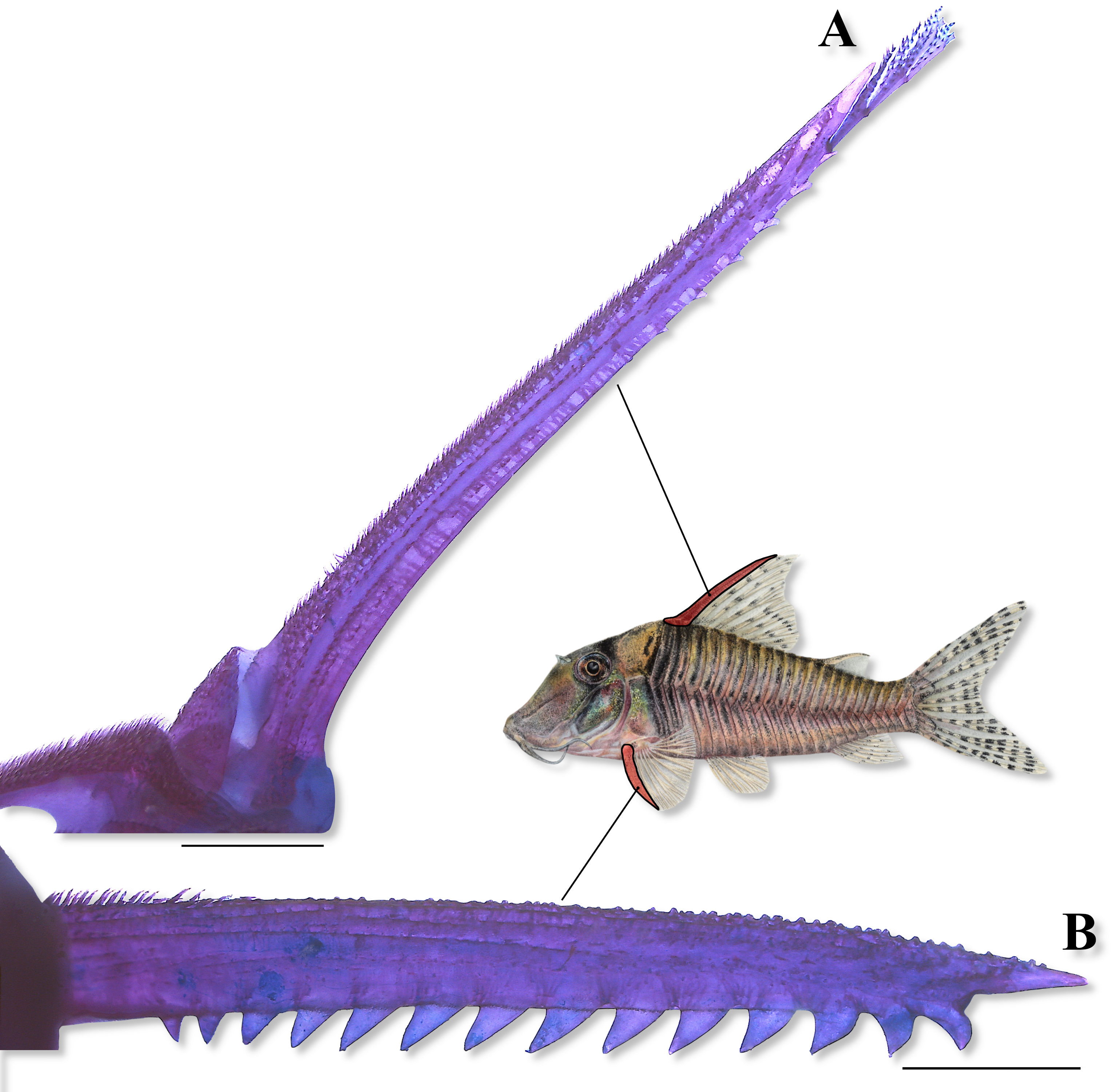
FIGURE 6| Lateral view of (A) the dorsal-fin spine and dorsal view of (B) the right pectoral-fin spine in a c&s paratype of Corydoras isaacisbrueckeri (CITL 1557, 43.0 mm SL), showing their serration patterns. Area where the illustrated bones are located in fish’s body marked in red in the miniature drawing of the new species. Scale bars = 1 mm.
Pelvic fin oblong, located just below second ventrolateral body plate, and at vertical through first or second dorsal-fin branched ray. Pelvic-fin rays i,5*(20); second branched ray typically as longest fin element, with rays decreasing in size towards both anterior and posterior margins of fin; first branched ray variably as longest fin element, or similar in size to second branched ray; last branched ray typically as shortest fin element, or unbranched ray in some specimens; unbranched ray with similar size to last branched ray in some specimens. Anterior internal process of basipterygium well developed and conspicuously laterally expanded, with dorsal lamina nearly vertically placed, ranging from well developed to conspicuously well developed, curved posteriorly and bent mesially; ventral laminar expansion obliquely placed, converging mesially towards anterior edge of process, conspicuously less developed than dorsal lamina; anterior external process laminar, typically moderately developed and slightly expanded posteriorly; dorsal ischiac process well developed, with anterior laminar expansion typically moderately expanded anteriorly, and posterior laminar expansion slightly expanded posteriorly; anterior and posterior laminar expansions of ischiac process roughly triangular; ventral ischiac process clearly smaller than dorsal process, roughly triangular, bent anteriorly (Fig. 7). Adipose fin roughly triangular, separated from base of last dorsal-fin ray by five to six dorsolateral body plates. Anal fin subtriangular, located just posterior to 12th or 13th ventrolateral body plates, and at vertical through adipose-fin spine base or anterior region of adipose-fin membrane. Anal-fin rays ii,5,i(7), ii,6*(11), i,7(2); third ray typically longest fin element, with rays decreasing in size towards both anterior and posterior margins of fin; fourth ray variably similar in size to third ray; last ray typically shorter fin element, or first ray. Caudal fin bilobed, with dorsal and ventral lobes similar in size or dorsal lobe slightly larger than ventral lobe; some specimens undergoing caudal regeneration with dorsal lobe smaller than ventral lobe. Caudal-fin rays i,12,i*(20), with generally five dorsal and ventral procurrent rays increasing in size posteriorly; small cartilage between upper principal and procurrent caudal-fin rays (presumably opisthural cartilage (Monod, 1968; McDowall, 1999)) observed in paratype CITL 1557, 40.9 mm SL (Fig. 8).
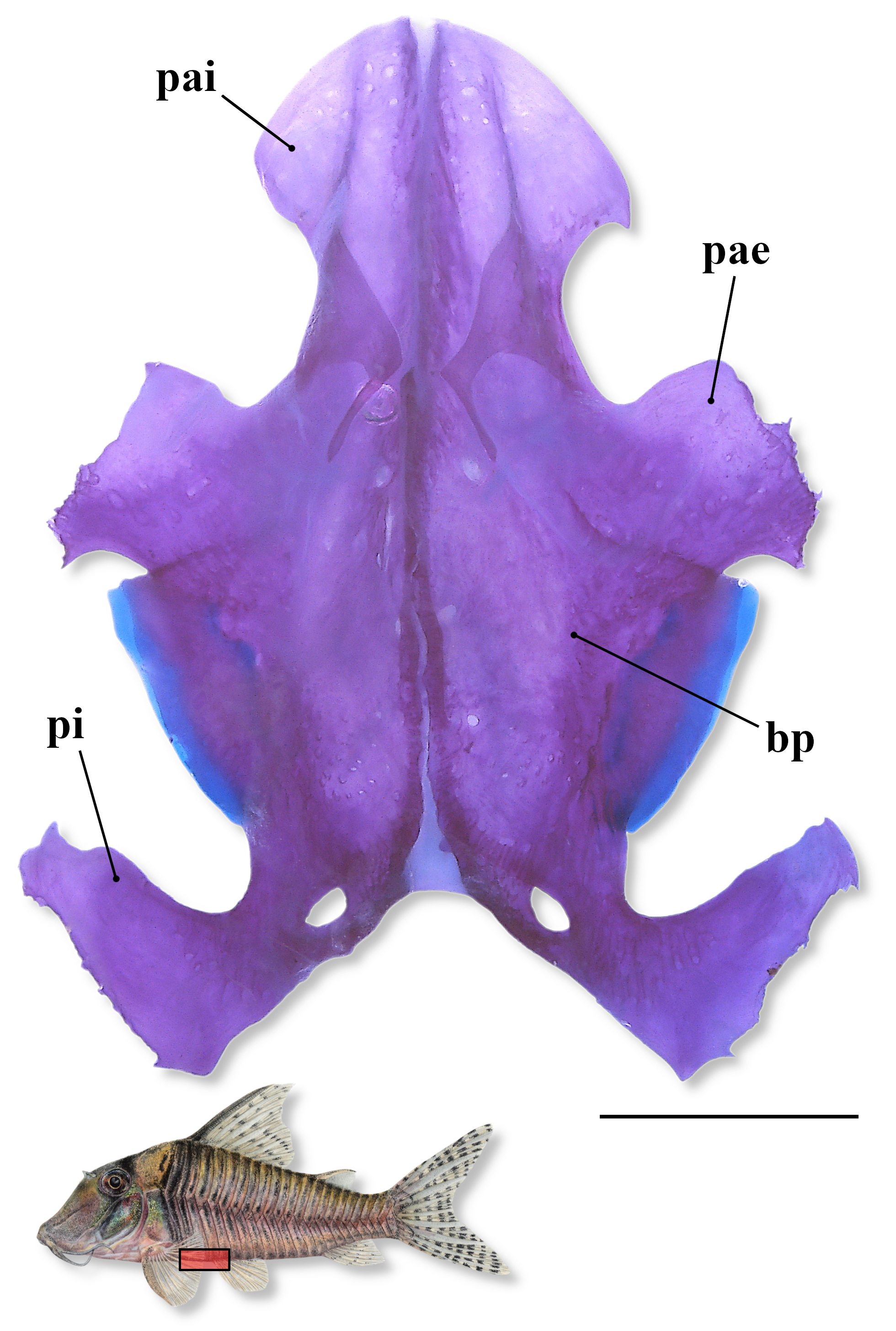
FIGURE 7| Pelvic girdle in a c&s paratype of Corydoras isaacisbrueckeri (CITL 1557, 43.0 mm SL). Abbreviations: bp: basipterygium, pae: anterior external process, pai: anterior internal process, pi: dorsal ischiac process. Area where the illustrated bones are located in fish’s body marked in red in the miniature drawing of the new species. Scale bar = 1 mm.
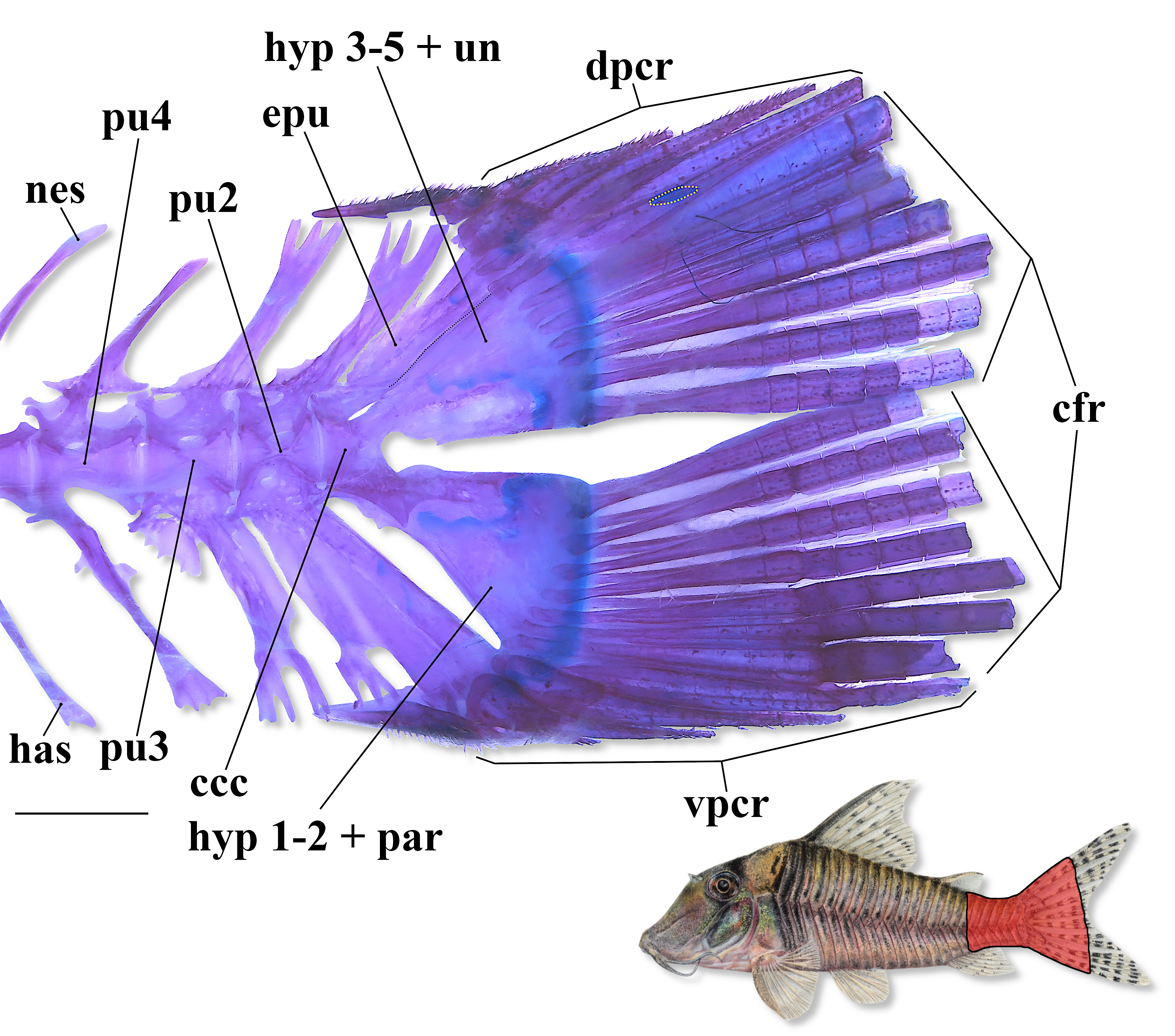
FIGURE 8| General morphology of caudal skeleton in a c&s paratype of Corydoras isaacisbrueckeri (CITL 1557, 40.9 mm SL), showing the small cartilage (yellow dotted line) between upper principal and procurrent caudal-fin rays. Abbreviations: ccc: compound caudal centrum, cfr: caudal-fin principal rays, dpcr: dorsal procurrent rays, epu: epural, has: haemal spine, hyp 1–5: hypurals 1 to 5, nes: neural spine, par: parhypural, pu 2–4: preural centra 2 to 4, un: uroneural, vpcr: ventral procurrent rays. Area where the illustrated bones are located in fish’s body marked in red in the miniature drawing of the new species. Scale bar = 1 mm.
Four to seven laterosensory canals on trunk; first ossicle tubular, second ossicle laminar, both bearing small odontodes; third, fourth, fifth, sixth and seventh, when present, encased in third, fourth, fifth, sixth and seventh dorsolateral body plates, respectively. Body plates with minute odontodes scattered over exposed area, with conspicuous line of odontodes confined to posterior margins. Dorsolateral body plates 23*(1), 24(18), 25(1); ventrolateral body plates 21*(19), 22(1). Dorsolateral body plates along dorsal-fin base 6*(8), 7(12); dorsolateral body plates between adipose- and caudal-fin 7*(1), 8(18), 9(1). Preadipose platelets 1(2), 2*(7), 3(10), 4(1). Ventral surface of trunk between posteroventral margin of cleithrum and pelvic-fin origin typically laterally delimited by first and second ventrolateral body plates, or only by first ventrolateral body plate; ventral portion of first ventrolateral body plate ranging from slightly to moderately expanded anteriorly. Small platelets covering base of caudal-fin rays; small platelets disposed dorsally and ventrally between junctions of lateral plates on posterior portion of caudal peduncle. Anterior margin of orbit, above region of junction between frontal and lateral ethmoid, region surrounding nasal capsule, and variably dorsal and lateral surfaces of snout with small-sized platelets bearing odontodes; platelets on snout, when present, scarce in c&s paratypes; some non-c&s paratypes with numerous odontodes on snout, suggesting snout covered by numerous small platelets. Ventral surface of trunk mostly covered by numerous small-sized platelets, except for region of pelvic girdle, which presents scarce platelets in both c&s paratypes; platelets irregular or roughly transversally elongated (relative to main axis of body) in shape and bearing odontodes (Fig. 9); in some non-c&s specimens, ventral surface of trunk on region of pelvic girdle and ventral surface of head with numerous odontodes, suggesting numerous small platelets covering both areas.
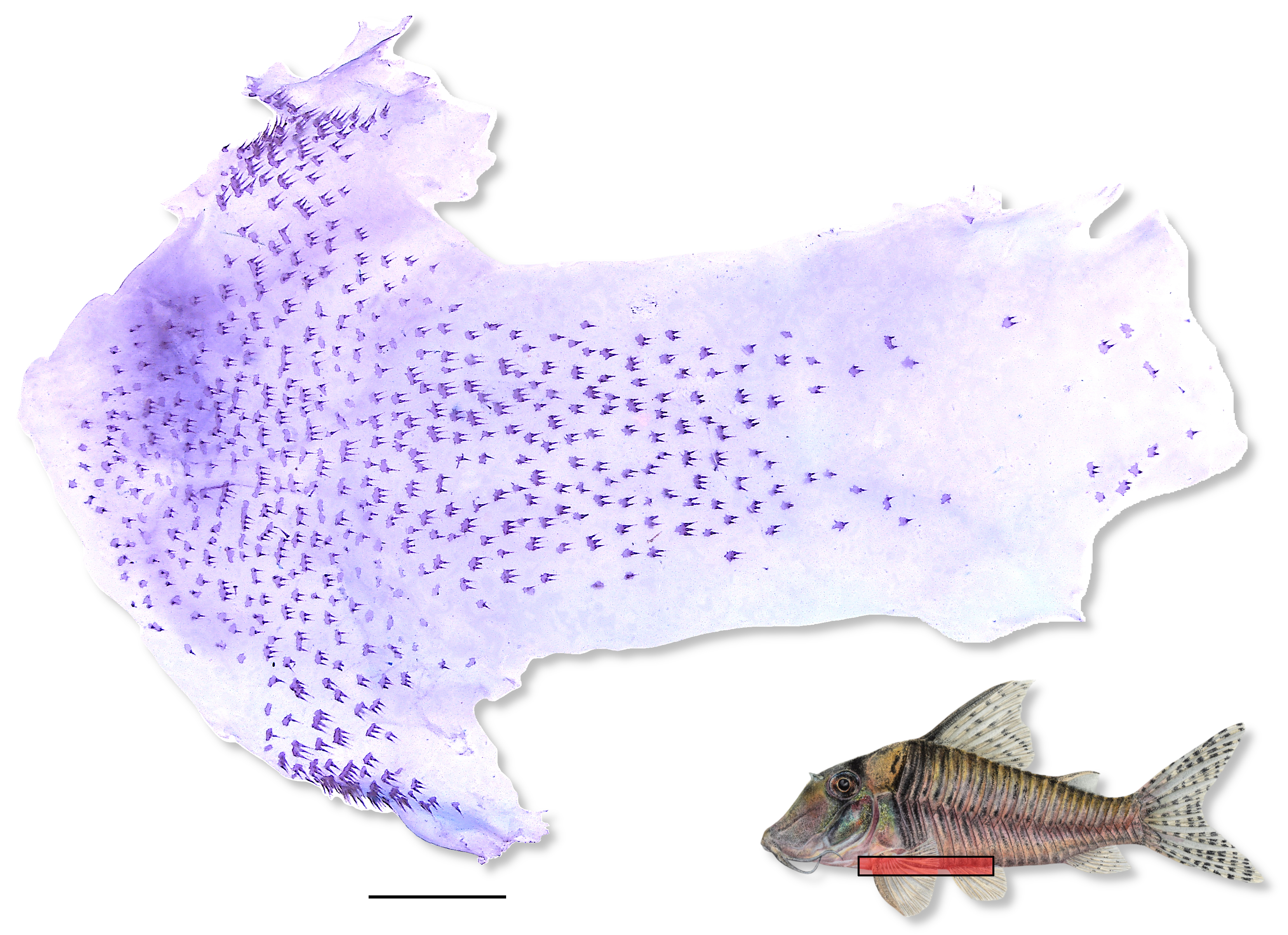
FIGURE 9| Ventral surface of trunk in a c&s paratype of Corydoras isaacisbrueckeri (CITL 1557, 43.0 mm SL), showing the small, irregular or roughly transversally elongated (relative to main axis of body) platelets. Area where the illustrated bones are located in fish’s body marked in red in the miniature drawing of the new species. Scale bar = 1 mm.
Vertebral count 22(2); ribs 5(2), first pair conspicuously large, its middle portion closely connected to first ventrolateral body plate; its tip not connected to anterior external process of basipterygium. Parapophysis of complex vertebra well developed.
Coloration in alcohol. Overall color pattern of body in Fig. 1. Ground color of body pale yellow or beige, with top of head dark brown. Dorsal and lateral surface of head densely covered by dark brown or black chromatophores, not forming smaller blotches; chromatophores conspicuously more concentrated dorsally and ventrally to orbit, forming large, elongated dark patch transversally crossing orbit, extending from anterior portion of parieto-supraoccipital towards region of interopercle, forming typical mask-like blotch; mask smoothly curved anteriorly to nearly straight; some specimens with conspicuous concentration of dark brown or black chromatophores on ventral half of opercle, forming irregular dark patch, typically fused with mask-like blotch; opercular patch variably diffuse; ventral portion of mask-like blotch just below ventral margin of orbit typically more evident and roughly arrowhead shaped; region just above posterodorsal margin of orbit slightly darker than dorsal portion of mask-like blotch, forming horizontally elongated eyebrow-like marking, variably nearly straight or slightly arched dorsally, following outline of orbit; eyebrow-like marking variably indistinct from mask-like blotch; region of contact between opercle and preopercle with conspicuous concentration of dark brown or black chromatophores; lips and barbels typically with dark brown or black chromatophores, conspicuously more concentrated on outer mental barbel; ventrolateral portion of head variably with dark brown or black chromatophores, with isthmus typically devoid of chromatophores. Cleithrum with dark brown or black chromatophores, especially on its laterodorsal surface, not forming small, dark blotches; chromatophores conspicuously more concentrated close to its posterior border, forming slender transversally elongated dark brown or black patch. First dorsolateral body plate covered by dark brown or black chromatophores, conspicuously more concentrated close to its posterior margin, forming slender transversally elongated dark patch. Region of contact between nuchal plate and posterior process of parieto-supraoccipital, middle portion of first dorsolateral body plate, and posterior process of parieto-supraoccipital clearly lighter than surrounding areas, forming roughly rounded pale area in dorsal view. Wide, transversally elongated dark brown or black patch extending from anterior portion of dorsal-fin base towards posteroventral margin of cleithrum, typically more evident until horizontal through middle portion of cleithrum; patch distinct but not solid, variably becoming gradually diffuse ventrally. Remaining dorso- and ventrolateral body plates covered by dark brown or black chromatophores, clearly more concentrated on middle portion of plates, typically forming slender, transversally elongated blotches; dark blotches variably fragmented into smaller, roughly rounded, irregular or vertically elongated blotches aligned in vertical rows along middle portion of lateral body plates; ventral portion of ventrolateral body plates, especially around pelvic fin, devoid of chromatophores or with fewer and/or less evident chromatophores; dark blotches typically becoming gradually diffuse ventrally and posteriorly. Posterior margin of body plates variably with conspicuous concentration of dark brown or black chromatophores, forming thin dark lines; dark lines on lateral series of plates typically becoming gradually diffuse towards ventral margin of flank. Dorsal fin with dark brown or black chromatophores, typically more concentrated on spine, branched rays, and along ventral margin of fin; branched rays with small, roughly rounded diffuse blotches, variably roughly aligned obliquely; some specimens with anterior portion of fin, on region between spine and second branched ray with more evident chromatophores, clearly darker than remaining portion of fin, but not forming conspicuous dark patch. Pectoral fin with dark brown or black chromatophores, clearly more concentrated on spine and rays, not forming small, dark blotches; chromatophores typically becoming gradually scarcer towards fin terminus, with last rays typically devoid of chromatophores. Pelvic fin devoid of or with scarce dark brown or black chromatophores. Anal fin with dark brown or black chromatophores, clearly more concentrated on rays and along fin base, typically forming small, faint dash-like dark blotches, especially on middle portion of fin; blotches roughly aligned vertically; blotches variably absent. Adipose fin with dark brown or black chromatophores, clearly more concentrated on spine, region of membrane close to spine, and along its base, typically not forming blotches; some specimens with small, roughly rounded to irregular faint dark blotches. Caudal fin with conspicuous concentrations of dark brown or black chromatophores, especially on rays, forming small, roughly rounded, irregular or horizontally elongated dark blotches, roughly aligned in transversal rows, forming slender bands; dark markings variably diffuse.
Coloration in life. Similar to color pattern of preserved specimens, but with brighter ground color of body (Fig. 10). Body covered by greenish yellow iridescent coloration, especially on posterior portion of head and cleithrum, forming large bright patches.
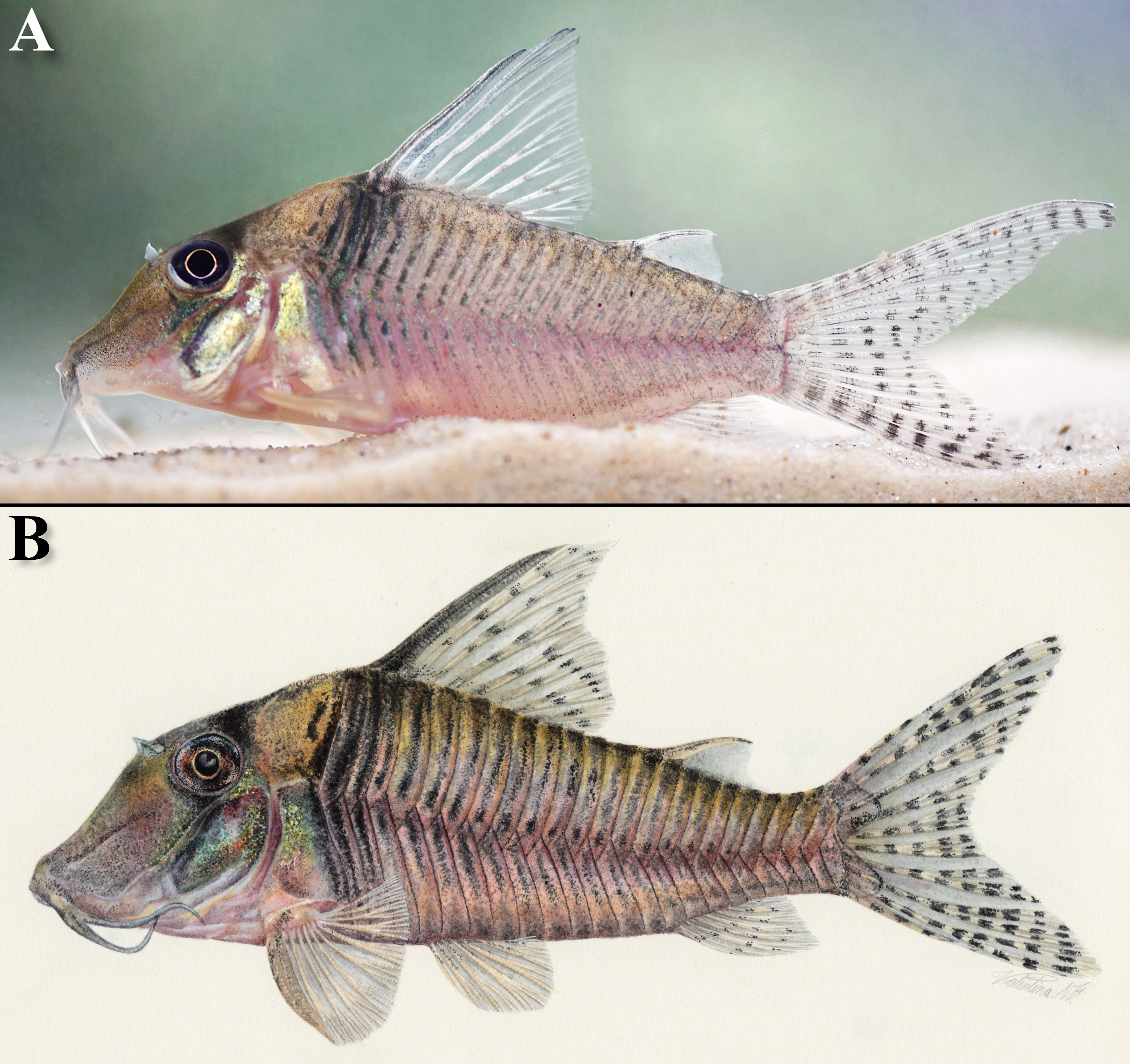
FIGURE 10| Color pattern in life of Corydoras isaacisbrueckeri in lateral view, showing (A) an uncatalogued specimen (unmeasured), and (B) a handmade drawing by Valentina Nieto based on the type specimens. Photo (A) by APH.
Sexual dimorphism. Males of Corydoras isaacisbrueckeri have a genital papilla, which is roughly lanceolate or tubular, a condition well-documented in Corydoradinae (see Britto, 2003; Nijssen, Isbrücker, 1980b; Spadella et al., 2017). Some specimens, most bearing a genital papilla, have first and variably second dorsal-fin branched rays elongated, a feature considered to be sexually dimorphic in some Corydoradinae species (e.g., Hoplisoma gryphus (Tencatt, Britto & Pavanelli, 2014) and H. longipinne (Knaack, 2007)). However, the presence of specimens lacking a tubular/lanceolate genital papilla (it is unclear if the absence of such a feature is due to any kind of damage on the region or not) but having elongated anterior dorsal-fin branched rays suggest that this feature cannot be considered sexually dimorphic in the new species. Moreover, elongated anterior dorsal-fin branched rays were observed in both juvenile and adult specimens. The presence of elongated anterior dorsal-fin branched rays in small/juvenile specimens has also been reported for C. iiap (see Tencatt et al., 2024b). Additionally, at least three male paratypes present a thickened pectoral spine, especially its distal half, which is generally covered by hypertrophied odontodes, similar to the illustrated by Nijssen, Isbrücker (1983:81, fig. 10g).
Geographical distribution. Corydoras isaacisbrueckeri is currently known from its type locality, the igarapé da ATM, with two additional records, the igarapé da Barreira and igarapé do Pavão, all right bank tributaries of the rio Tefé, itself a tributary of the right bank of the rio Solimões, Amazonas State, Brazil (Fig. 11).

FIGURE 11| Map showing the known geographic records of Corydoras isaacisbrueckeri (red star: type locality, the igarapé da Associação dos Trabalhadores do Mamirauá plus the igarapé do Pavão; black diamond: igarapé da Barreira), Amazonas State, rio Tefé basin, Brazil.
Ecological notes. The three collecting sites of Corydoras isaacisbrueckeri are clear water streams located in areas of “terra firme”, with an average of around 7.0 m width, 50.0 cm deep, and low water current. The streams have relatively large riparian forest areas, with only part of their margins deforested for subsistence agricultural plantations (Fig. 12). Representatives of the new species were captured only on marginal sandbanks (small beaches), with coarse, yellow sand. The following physical-chemical water parameters were obtained at the igarapé da ATM (Fig. 12A) during the dry season of 2024 (August to October): water temperature 25.6° C, pH 6.2, conductivity 45.6 µS/cm3, and dissolved oxygen 4.78 mg/L. Although more than 60 other fish species were collected with the new species, no syntopic Corydoradinae species were found (APH, pers. obs.).

FIGURE 12| Natural habitat of Corydoras isaacisbrueckeri, showing (A) the igarapé da Associação dos Trabalhadores do Mamirauá, (B) the igarapé da Barreira, and (C) the igarapé do Pavão, all tributaries of the rio Tefé, Amazonas State, Brazil.
Etymology. Corydoras isaacisbrueckeri is named in honor of Dr. Isaäc Jan Hendrik Isbrücker, beloved friend and renowned Loricarioidea expert. Isaäc devoted most of his life to the aquarium hobby and ichthyology, publishing dozens of scientific articles on Corydoradinae and Loricariidae along his career. This small homage intends to highlight how special Isaäc is as a human being, as well as a devoted scientist, inspiring LFCT on personal and professional levels. A noun in apposition.
Conservation status. Currently, the new species is known only from its type locality, the igarapé da ATM, plus two additional records, the igarapé do Pavão and igarapé da Barreira, all tributaries of the rio Tefé in Amazonas State, Brazil. The Conservation Unit Tefé Floresta Nacional (FLONA) is a part of the Corredor Ecológico Central da Amazônia (Central Amazon Ecological Corridor) in Western Amazon, which, in the middle rio Solimões region, is a part of the Federal Conservation Units located in the Tefé Region and the Reserva Extrativista (RESEX) of the middle rio Juruá, in addition to State Conservation Units, namely: the Reserva de Desenvolvimento Sustentável (RDS) of Mamirauá, RDS Amanã, RDS Catuá Ipixuna, and RDS Cujubim. Additionally, it is important to mention that the middle rio Solimões region is relatively distant from the major deforestation frontiers, presenting relatively low external pressure from the affected areas. Nonetheless, drought and rising temperatures have led to large events of fish mortality in several regions of the Amazon, including in the lago Tefé and its tributaries. In this context, the environmental impacts due to climate change (e.g., Marengo et al., 2024; Marmontel et al., 2024) are surely increasing the risk of extinction of the amazonian aquatic species. However, considering that the new species occurs in a relatively well-preserved region within the Brazilian Amazon, it is likely that such negative impacts currently do not represent a threat to the species as whole. Therefore, considering the currently available data and according to the International Union for Conservation of Nature (IUCN) categories and criteria (IUCN Standards and Petitions Subcommittee, 2024), Corydoras isaacisbrueckeri would be classified as Least Concern (LC).
Remarks. In the aquarium trade, at least two putative undescribed species are currently recognized under the codes Corydoras sp. CW128 (Fig. 13A), from the quebrada Zaragoza, a tributary of the rio Marañon in Peru (Hans Evers, 2025, pers. comm.), and CW207 (Fig. 13B), of which the locality is only referred to as “Colombia”. Besides occurring in the same drainage system, as the rio Pastaza is also a tributary of the rio Marañon, the color pattern of Corydoras sp. CW128 resembles the one of C. pastazensis, especially when compared to some paratypes with smaller blotches along midline of flank (Fig. 14A), while the new species lacks such distinct series along the midline of flank, and is devoid of moderate-sized, dark blotches on flanks posteriorly to the large dark patch below dorsal fin (Figs. 1, 10). Additionally, CW128 also has the large, vertically elongated dark patch below dorsal fin intensely pigmented, which is different from the more diffuse dark patch below dorsal fin in C. isaacisbrueckeri. The smaller, dark blotches on flanks of CW128 are more evident and roughly aligned in longitudinal rows, especially close to the midline of flank on anterior portion of flanks, which is more similar to the observed in the type specimens of C. pastazensis, although the dark markings on the latter are clearly larger. In the new species, the blotches are clearly scarcer and less evident, not forming longitudinal rows in any portion of flank.

FIGURE 13| Uncatalogued aquarium specimens of (A) Corydoras sp. CW128, and (B) Corydoras sp. CW207, showing their general color pattern in life in lateral view. Photo (A) by Hans Evers, and (B) by Manyork Chow.

FIGURE 14| General morphology and color pattern in lateral view in (A) a paratype of Corydoras pastazensis (USNM 164464, unmeasured), and three paratypes of Corydoras pastazensis orcesi, with (B) specimen USNM 203827, unmeasured, (C) BMNH 1970.4.17.3–4, 56.6 mm SL, and (D) ZMA 110.377, 50.6 mm SL. Photo (A) by MRB, (B) by Sandra Raredon, (C) and (D) by SAS.
Considering this, it seems that Corydoras sp. CW128 corresponds to the morphotype of C. pastazensis from the rio Ucayali basin illustrated by Nijssen, Isbrücker (1986:59, fig. 42). Interestingly, the analysis of a photograph of a single adult specimen from the rio Ucayali basin (NRM 28578) showed a large and conspicuous dark patch on the anterior portion of the dorsal fin, which was not observed in any of the examined specimens of C. isaacisbrueckeri nor in typical C. pastazensis. In relation to CW207, besides the vague locality, our analysis was restricted to photos of a single specimen (Fig. 13B), in which the vertically elongated dark patch below dorsal fin is intensely pigmented, as in CW128, whereas the remaining dark markings on flanks are faint, nearly imperceptible, although what seems to be a longitudinal series of dark blotches along the midline of flank can be observed by zooming the image. Therefore, this species can also represent a form of C. pastazensis, but this is something that needs to be further investigated considering the scarce photographic material.
Corydoras pastazensis was described based on four specimens from the rio Bobonaza, a tributary of the rio Pastaza in Ecuador. According to Weitzman (1963), the species has a dark bar below dorsal fin (reported to be diffuse on the holotype), and moderate-sized dark blotches irregularly set on remaining portions of flanks. Although widely recognized as a masked species, Weitzman (1963) made no mention to this feature, only stating that the anterior portion of the head, which he considered to be “that portion exclusive of the opercula”, i.e., from the tip of snout to region just anterior to the opercle, is grayish brown. Weitzman, Nijssen (1970) proposed a subspecies to C. pastazensis, C. pastazensis orcesi, based on 21 specimens captured in tributaries of the rio Tigre, Ecuador. Both the rio Pastaza and rio Tigre are tributaries of the rio Marañón, which flows into the rio Amazonas. The authors used the color pattern to differ C. p. orcesi from C. p. pastazensis, as the first presents (I) the posterior border of the vertical dark blotch below dorsal fin convex (vs. concave in C. p. pastazensis), and (II) two moderate-sized blotches along flank midline, the first just below the dorsal-fin posterior margin and the second just below the adipose fin (vs. absence of such blotches on flank midline, which only presents “small spots and very pale blotches”). Weitzman, Nijssen (1970) also mentioned that C. p. orcesi possesses a dark mask-like blotch and, although never reported to be present in C. p. pastazensis, such feature was never proposed as diagnostic in that or in subsequent works.
Nijssen, Isbrücker (1986) analyzed 44 specimens, including the type-series of C. pastazensis and of C. p. orcesi, adding specimens from the rio Napo in Ecuador, and rio Ucayali systems. The authors described the color pattern of these additional populations, highlighting the difference in color pattern when comparing the population from the rio Ucayali basin with those from the Napo, Pastaza, and Tigre drainages. In the specimens from the rio Ucayali basin, the vertical blotch below dorsal fin is narrow (vs. wide), and the flank is densely covered by smaller, conspicuous dark blotches, which are roughly vertically aligned but also roughly aligned in longitudinal rows, especially close to the flank midline on anterior portion of trunk, with only a few larger blotches on the flank (vs. flanks with scarce, moderate-sized blotches). Even considering these differences, the population from the rio Ucayali was considered as conspecific with the others. In this context, Nijssen, Isbrücker (1986) placed C. p. orcesi in the synonymy of C. pastazensis.
Regardless of the synonymy proposed by Nijssen, Isbrücker (1986), some authors continued to use the name C. orcesi (e.g., Alexandrou et al., 2011; Lima, Sazima, 2017). Such decision may have been influenced by Isbrücker’s (2001) catalog of the genera and species of Corydoradinae, which considered all nominal species of Corydoradinae as potentially valid. Moreover, the alleged differences in color pattern provided in the original description likely reinforced this idea, as the presence of two moderate-sized dark blotches along flank midline was reported only for C. orcesi (Weitzman, Nijssen, 1970), something that was not contested in Nijssen, Isbrücker (1986). Contrary to Weitzman (1963), Weitzman, Nijssen (1970), and Nijssen, Isbrücker (1986), the analysis of the holotype (USNM 177216) and the three paratypes (USNM 164464) of C. pastazensis revealed that this species presents the two moderate-sized dark blotches longitudinally aligned on the flank midline, one just posterior to posterior margin of dorsal fin (or below region of the first preadipose platelet), and the other one below adipose fin, typically in a vertical with middle to posterior portion of adipose fin (Figs. 14A, 15A). These blotches are placed in the exact same areas as those of the holotype (USNM 204358) and examined paratypes (BMNH 1970.4.17.3–4, USNM 203827, 203828, ZMA 110.377, 110.378, and 110.379) of C. p. orcesi (Figs. 14B–D, 15B).

FIGURE 15| General morphology and color pattern in lateral view in (A) the holotype of Corydoras pastazensis (USNM 177216, 46.2 mm SL), and (B) the holotype of Corydoras pastazensis orcesi (USNM 204358, 52.9 mm SL). Photos by Sandra Raredon.
In both type series, it was possible to note that the size of these blotches is slightly variable among the specimens, with some of them presenting smaller and others larger blotches, although always arranged in a distinct longitudinal series along the flank midline (Figs. 14, 15). Moreover, the number of these midlateral blotches is also variable among the type specimens, ranging from two to four blotches (Figs. 14, 15), a number that can be even higher considering aquarium specimens, which can have up to six blotches (LFCT, pers. obs.). The blotch on posterior portion of caudal peduncle, when visible, is clearly more diffuse than remaining midline blotches (Figs. 14, 15), which may be the reason why some specimens are considered to present only two midlateral blotches instead of three, as the holotype of C. p. orcesi (Fig. 15B). Additionally, the peduncular blotch is conspicuously vertically elongated, forming a relatively narrow, diffuse dark bar covering the posterior portion of the caudal peduncle, differing from the roughly rounded or narrow, slightly vertically elongated shape of the remaining midlateral blotches (Figs. 14, 15). Considering how faded these specimens are now, it is uncertain if the peduncular blotch is always present. In any case, we still consider that some specimens present only two midlateral blotches based on the current state of the examined specimens.
Regarding the presence of a dark mask-like blotch, we were able to observe a diffuse mask-like blotch only in part of the paratypes of C. p. orcesi. In the holotypes of both taxa, it was possible to observe more concentrated dark brown or black chromatophores ventrally to the orbit (more evident on the holotype of C. p. orcesi), which is likely a remnant of a mask-like blotch. Overall, the dark pigmentation of the holotype of C. pastazensis is clearly more faded than that of the holotype of C. p. orcesi, which explains different interpretation of both color patterns by previous authors (i.e., Weitzman, 1963; Weitzman, Nijssen, 1970; Nijssen, Isbrücker, 1986). As for the shape of the large, dark vertically elongated blotch below dorsal-fin anterior portion, it was not possible to establish any difference between the specimens of both type series, as such feature seems to be quite variable, with specimens presenting posterior margin of this blotch roughly concave, convex or even straight. Nonetheless, even diffuse, the color pattern of the holotype of C. pastazensis is clearly compatible with that of C. p. orcesi (Fig. 15), corroborating the synonymy proposed by Nijssen, Isbrücker (1986).
In Alexandrou et al. (2011), however, C. pastazensis and C. orcesi appear as two distinct species, which was used in previous articles as an argument to consider the latter as valid (see Lima, Sazima, 2017). Although the origin of the voucher specimens (two of C. pastazensis and one of C. orcesi) of both species was stated as “Amazon”, the LBP database showed that these vouchers are aquarium specimens purchased in England. The two identified as C. pastazensis in Alexandrou et al. (2011) have a single voucher specimen each, deposited in the LBP collection. The single voucher specimen identified as C. orcesi is not deposited in LBP, which holds only a single tissue sample of this specimen. We examined the specimens attributed to C. pastazensis (LBP 2835 and 2836; Figs. 16A, B) by Alexandrou et al. (2011), together with photographs taken closer to the time of preservation (Figs. 16C, D). Our analysis revealed that none of these specimens seem to represent C. pastazensis. Although faded, the specimen LBP 2835, 57.8 mm SL (Figs. 16A, C) have an eye mask, a vertically elongated dark patch below the anterior portion of dorsal-fin base, small dark markings on the flanks, and the anterior portion of the dorsal fin with a conspicuously dark patch, resembling the color pattern of Corydoras sp. CW128. The specimen LBP 2836, 41.7 mm SL (Figs. 16B, D) have an eye mask, a roughly rounded dark patch below the anterior portion of the dorsal-fin base, and small, dark blotches on flanks, resembling C. blochi. Although the aquarium specimen attributed to C. orcesi by Alexandrou et al. (2011) was not examined herein, it is clear that the specimens LBP 2835 and LBP 2836 are not compatible with C. pastazensis,and, consequently, with C. orcesi, as they were confirmed as conspecific herein. Therefore, the argument that the results of Alexandrou et al. (2011) confirmed the validity of C. orcesi becomes unfounded.

FIGURE 16| General morphology and color pattern of the voucher specimens attributed to Corydoras pastazensis in Alexandrou et al. (2011), showing specimens (A, C) LBP 2835, 57.8 mm SL, and (B, D) LBP 2836, 41.7 mm SL, in lateral view. Photos (A) and (B) by SAS, and (C) and (D) by Claudio Oliveira.
Discussion
Different from other genera within Corydoradinae, such as Aspidoras Ihering, 1907, Brochis, Hoplisoma, and Scleromystax Günther, 1864, Corydoras presents a quite conservative morphology, with all species showing overall similar shape and osteological features. In this context, differences in color pattern are extremely valuable in the recognition of the species in Corydoras. Phylogenetically, Corydoras forms two major clades, a smaller one harboring C. coriatae Burgess, 1997, C. fowleri Böhlke, 1950, and C. semiaquilus Weitzman, 1964, and a larger one including the remaining species (Alexandrou et al., 2011; Dias et al., 2025). Morphologically, the only difference between the species of these two clades is the presence of small- to relatively large-sized coalescent platelets, forming a typical mosaic-like pattern on ventral surface of head and trunk (vs. platelets on ventral surface of head and trunk, if present, small, not coalesced). The presence of these coalesced platelets, especially on ventral surface of trunk, is independently shared with at least two other genera within Corydoradinae, Brochis and Hoplisoma (see Tencatt, Britto, 2016; Tencatt et al., 2019, 2023b). Interestingly, events of convergence in Corydoradinae occurred not only in color pattern traits (see Alexandrou et al., 2011), but also in some morphological traits, as discussed in Tencatt et al. (2025a,c).
Some of these homoplastic features are potentially paedomorphic, such as the absence of contact between the parieto-supraoccipital and the nuchal plate, which occurs in nearly all genera within Corydoradinae, with Corydoras as the only exception. It is important to mention that not all species of Corydoras were examined herein, so, such feature may eventually be found in this genus in future analysis. Other interesting potentially paedomorphic feature refers to the presence of a distinct longitudinal series of dark blotches along flank midline, which is present in C. pastazensis. Many Corydoradinae displays the flank midline series of blotches in larval/juvenile stage (see Fuller, 1999, 2012), which eventually disappears or is even modified (e.g., blotches coalesce and turn into a stripe) during the individual’s growth (see Tencatt, Evers, 2016; Tencatt et al., 2021, 2023b, 2024a,b), while other groups retain such feature in adult stage (e.g., Tencatt et al., 2013, 2014a,b, 2016, 2022b, 2025a).
Therefore, the presence/absence of a distinct series of blotches along flank midline in adult specimens should not be treated as a color pattern variation, but rather the presence/absence of a putative paedomorphism. In this context, although overall similar in morphology (like all congeners), we consider that the distinct longitudinal series of at least two dark blotches along flank midline in C. pastazensis, which is absent in C. isaacisbrueckeri,represents a robust distinguishing feature between them. At the same time, the sharing of a distinct longitudinal series of dark blotches along the midline of flank (among other features; see above discussion), present in both holotypes of C. pastazensis and C. p. orcesi, represents solid evidence that they are indeed conspecific. Regardless of the identity of Corydoras sp. CW128 (i.e., whether or not it is conspecific with C. pastazensis), we still consider its color pattern as different from the one displayed by the new species, not only by the distinct longitudinal series of dark blotches along flank midline (absent in the new species), but also by the arrangement and/or intensity of pigmentation of the dark markings on the flanks in CW128 (see Remarks section; compare Fig. 10A with Fig. 13A).
While comparing the different snout shapes in Corydoradinae, Tencatt et al. (2025a) also noted some interesting differences in the morphology of the mouth and barbels, which will be similarly explored hereafter. For practical and standardization purposes, we summarized the mouth-related structures analyzed herein in Tab. 2, which were all depicted herein (Figs. 3, 17–19). Britto (2003) analyzed only two characters related to the external morphology of mouth/barbels: (char. 79) area at the corner of mouth, ventral to the maxillary barbels smooth (state 0), or with a fleshy flap (state 1); and (char. 80) lower lip as a plain skin fold, lacking inner mental barbel (state 0), or with a single inner mental barbel, the latter character already analyzed in Reis (1998). Ten years later, Vera-Alcaraz (2013) broadened the analysis of mouth/barbels morphology, considering four characters, as follows: (char. 191) shape of the flap expansion of the lower lip, which can be small, restricted laterally (state 0), small, wholly bordering mouth (state 1), small, wholly bordering mouth and notched mesially (state 2), or broad, wholly bordering mouth (state 3); (char. 192) shape of the inner mental barbel (referred as “mesial barbel”), which can be absent (state 0; sic), one conical (state 1), one flat (state 2), and two flat (state 3); (char. 193) length of maxillary barbel, which can be short (state 0), long (state 1), or extremely long (2), as proposed in Reis (1998; see char. 68); and finally character 194, area at the corner of mouth, ventral to the maxillary barbels smooth or with a very shallow ridge of skin (state 0), or with a distinct flap of skin (state 1).
TABLE 2 | Characters related to the mouth and their respective putative character-states (not polarized), all raised herein.
Characters | Character-states | |||
| (0) | (1) | (2) | (3) |
1. Lateral fold at anteroventral portion of snout, length | Long, reaching or surpassing the anterior margin of the orbit | Short, not reaching the anterior margin of orbit, |
|
|
2. Lateral fold at anteroventral portion of snout, delimitation of lateral margin | Delimited by lateral ethmoid and infraorbital 1 | Entirely fleshy, not delimited by bony structures | Delimited by the ventral margin of infraorbital 1 | Delimited by the preinfraorbital bony plate and ventral margin of infraorbital 1 |
3. Maxillary bulb, dorsolateral skin fold | Absent | Present |
|
|
4. Maxillary bulb, posteroventral margin | With conspicuous roughly rounded to triangular fleshy expansion | Smooth, just slightly projected laterally and/or ventrally |
|
|
5. Lower lip, mesial notch | Deep, with an acutely angled V-shaped contour between each inner mental barbel | Shallow, with a slightly rounded or mild angled V-shaped contour between each inner mental barbel |
|
|

FIGURE 17| Detail of the mouth and barbels in a c&s specimen of Hoplisoma sp. CW127 (CITL 1572, 46.3 mm SL), in (A) lateral and (B) ventral views. Abbreviations: imb: inner mental barbel, ll: lower lip, lr: labial ramus, mba: maxillary barbel, mar: maxillary axial rod, mr: mental ramus, mx: maxilla, omb: outer mental barbel, ul: upper lip. Red arrows indicate the branches of the labial ramus at each inner mental barbel base. Area where the illustrated structures are located in fish’s body marked in red in the miniature photo of an uncatalogued specimen of Hoplisoma sp. CW127 in life (photo by Willian Ohara). Scale bars = 1 mm.
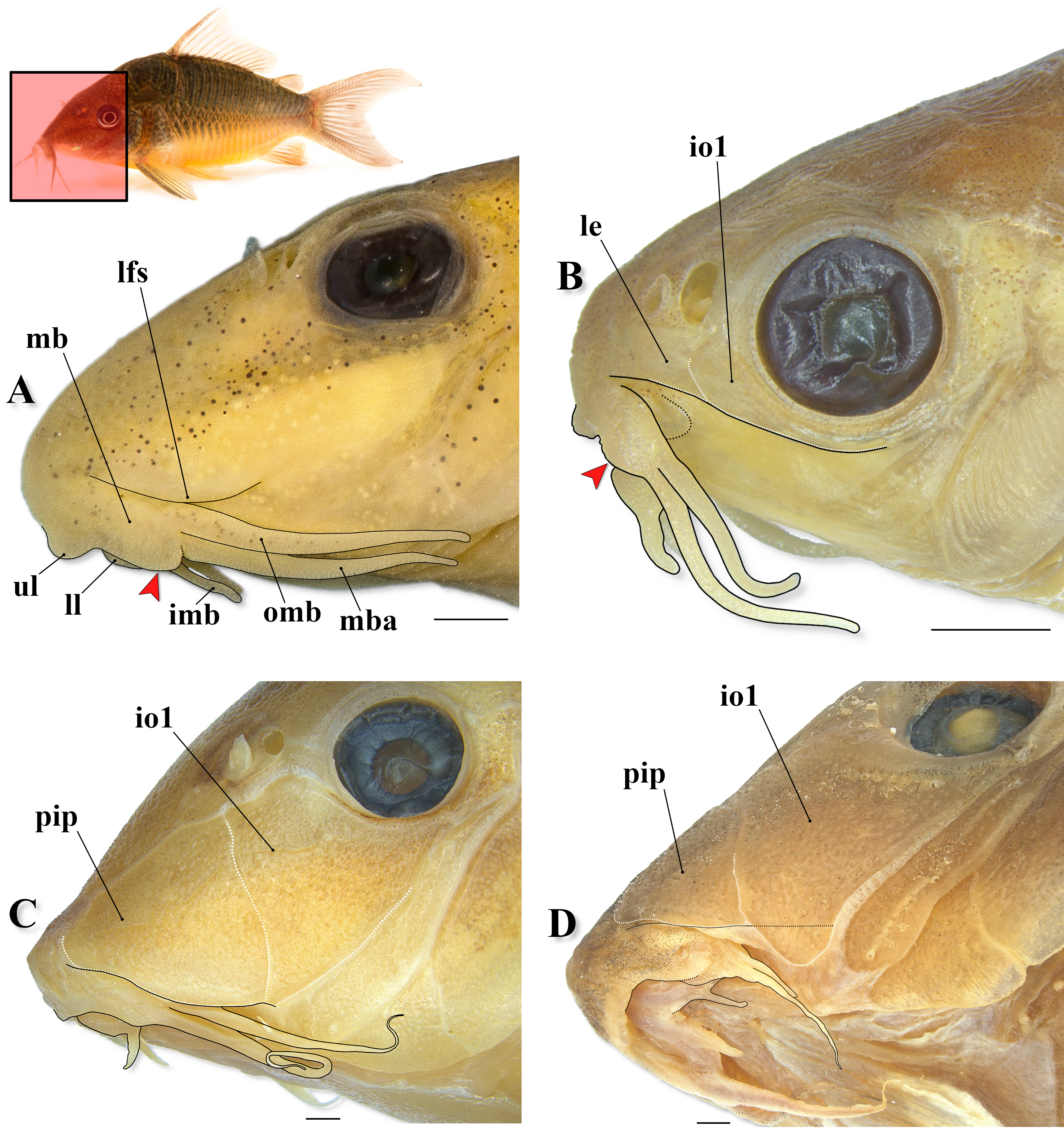
FIGURE 18| Detail of the snout and mouth in some Corydoradinae, showing the mouth-related structures (outlined in black) in (A) the holotype of Aspidoras psammatides (MNRJ 28407, 25.7 mm SL), (B) Gastrodermus hastatus (MNRJ 54328, 20.7 mm SL), (C) Brochis britskii (LBP 688, 60.5 mm SL), and (D) Brochis cf. splendens (CPUFMT 3792, 69.0 mm SL). Abbreviations: imb: inner mental barbel, io1: infraorbital 1, le: lateral ethmoid, lfs: lateral fold at anteroventral portion of snout, ll: lower lip, mb: maxillary bulb, mba: maxillary barbel, omb: outer mental barbel, pip: preinfraorbital plate, ul: upper lip. White dotted line delimiting lateral ethmoid and infraorbital 1 in (B), and preinfraorbital plate and infraorbital 1 in (C) and (D). Red arrows in (A) and (B) indicate the conspicuous posteroventral fleshy expansion of the maxillary bulb. Area where the illustrated structures are located in fish’s body marked in red in the miniature photo of Brochis sp. in life (photo by Martin Taylor). Scale bars = 1 mm.

FIGURE 19| Detail of the snout and mouth in some Corydoradinae, showing the mouth-related structures (outlined in black) in (A) Osteogaster rabauti (LBP 2827, 36.6 mm SL), and (B) in the holotype of Aspidoras azaghal (MNRJ 51793, 33.4 mm SL), in lateral and ventral views, respectively. Abbreviations: imb: inner mental barbel, ll: lower lip, mb: maxillary bulb, mba: maxillary barbel, omb: outer mental barbel, ul: upper lip. White dotted line in (A) delimiting infraorbital 1, and red square in (B) highlighting the region of the mesial notch of the lower lip. Area where the illustrated structures are located in fish’s body marked in red in the miniature photo of Osteogaster eques in life (photo by Willian Ohara). Scale bars = 1 mm.
Britto’s (2003) character 80 and Vera-Alcaraz’s (2013) character 193 will not be addressed in this discussion as all representatives of Corydoradinae possess a pair of inner mental barbels, and relatively short maxillary barbel, being, therefore, uninformative in the context of this work (i.e., phylogenetic relationships among Corydoradinae). In relation to Vera-Alcaraz’s (2013) character 193, the author mentioned that some Aspidoras species have a long maxillary barbel, reaching to region of pectoral-fin insertion. Considering our examined material (see Oliveira et al., 2017; Tencatt, Bichuette, 2017; Tencatt et al., 2020, 2022b), the maxillary barbel in Aspidoras, at most, slightly surpasses anteroventral limit of gill opening, not reaching pectoral-fin origin. Similarly, the character 191 in Vera-Alcaraz (2013) will also be omitted in this discussion because the lower lip reduced to a small flap, entirely bordering the mouth and with a medial notch (state 2) is shared by all Callichthyidae. Nonetheless, in Corydoradinae, it is possible to observe differences related to the extension of the mesial notch on lower lip among representatives of the different genera, and within the same genus, such as in Brochis. This character (i.e., extension of the mesial notch of the lower lip), which was raised herein, will be addressed below.
As aforementioned, the peculiar structure at the corner of the mouth (i.e., Britto’s, 2003, char. 79), more often referred to as “fleshy flap”, was widely used in the literature to recognize species within Corydoras (see data compiled in Dias et al., 2025). Curiously, Vera-Alcaraz (2013:94), stating that his character 194 corresponds to Britto’s (2003) character 79, coded such condition as present (state 1) only for C. cervinus Rössel, 1962. In his character’s 194 description, Vera-Alcaraz (2013:94) indicated that such condition was only considered present in specimens where this fleshy flap is elongated, with a barbel-like aspect, also known as the “third rictal barbel” (see Nijssen, 1970). The presence of elongated posteriorly fleshy flap is indeed uncommon in Corydoras (see Tencatt et al., 2020, 2021, 2024a,b), which would explain why the author found this feature present only in a single Corydoras species. In any case, it is clear that Britto’s character 79 and Vera-Alcaraz’s (2013) character 194 are not the same as the authors made different interpretations.
Recently, Tencatt et al. (2025a:11, fig. 3) mentioned the presence of a wrinkle of skin in both Hoplisoma noxium Tencatt, Ohara, Carvalho, Grant & Britto, 2025 and H. tenebrosum Tencatt, Ohara, Carvalho, Grant & Britto, 2025, which likely represents what Vera-Alcaraz called of “very shallow ridge of skin”. Such structure is somewhat similar to that observed in Corydoras, but differs from it by being apparently composed only of skin, differing from the fleshy (thicker), roughly triangular (or conical when laterally abducting lateral portion of mouth) tubercular structure found in Corydoras. Moreover, in the species with a wrinkle of skin (i.e., a flat skin fold) at the corner of the mouth, such region becomes typically smooth when laterally abducting the maxillary bulb. Alternatively, some species have a larger wrinkle of skin at the mouth corner (e.g., Scleromystax barbatus (Quoy & Gaimard, 1824),and some specimens of Aspidoras psammatides Britto, Lima & Santos, 2005 and Gastrodermus hastatus (Eigenmann & Eigenmann, 1888)), in which a shallow, flat wrinkle of skin is still perceptible even when laterally abducting the maxillary bulb. However, it is important to mention that only through a histological analysis can this assumption be undoubtedly confirmed. Even so, we consider that, although in the same area, the different external morphology of both structures suggest that they are not homologous.
In any case, the presence of a wrinkle of skin at the mouth corner is the typical condition in Corydoradinae, with few representatives having the corner of mouth presumably smooth (with mouth retracted), such as some species within the Brochis reticulata (Fraser-Brunner, 1938) and B. splendens groups sensu Tencatt et al. (2025a), and some Gastrodermus (e.g., G. guapore (Knaack, 1961), G. undulatus (Regan, 1912)). In those groups, the maxillary bulb tends to be closely attached to snout, making it difficult to see the area at the corner of the mouth, as they typically have deeper ventrolateral groove just ventral to the anterior portion of the lateral fold at the anteroventral portion of the snout, where the maxillary bulb fits closely. In most Corydoradinae (except for Corydoras), some specimens are often preserved with tightly retracted mouth, making the maxillary bulb closely attached to anterior portion of the lateral fold, making it difficult or even impossible to observe if the wrinkle of skin is indeed present (with normally retracted mouth), as the corner of the mouth becomes smooth when laterally abducting the maxillary bulb. In Aspidoras, the visualization of the corner of the mouth is even more challenging, as the anterior portion of the lateral fold tends to be projected ventrally towards the maxillary bulb, typically covering its anterodorsal portion. Therefore, it is uncertain if these species indeed present the lateral corner of the mouth smooth or if the wrinkle of skin is just too reduced and/or hidden below the maxillary bulb.
Finally, Vera-Alcaraz’s (2013) character 192, which refers to the shape of the inner mental barbel, is described with four character-states: inner mental barbel absent (state 0), with one conical barbel (state 1), or one (state 2) or two (state 3) flat barbels. Interestingly, the author considered two distinct traits, i.e., the number, which is basically Britto’s (2003) character 80, and shape of the inner mental barbel, as part of a single character. Firstly, regarding the number of barbels, we consider that the presence of inner mental barbel in Callichthyinae is questionable. In most representatives, except for most Callichthys Scopoli, 1777, Hoplosternum Gill, 1858 and variably Megalechis Reis, 1997, which present smooth lower lip, the lower lips seem to be notched, one (in Dianema Cope, 1871, Lepthoplosternum, and variably Megalechis) or multiple (Callichthys serralabium Lehmann A. & Reis, 2004) times, forming roughly triangular to rounded flat projections in their lower lips. Contrary to the conical inner mental barbel in Corydoradinae, which presents a distinct base emerging from the lower lip, these flat labial expansions are indistinct from the remaining portions of the lower lip, suggesting that they resulted from notching in the labial fold itself. Considering this, it seems that these labial expansions in Callichthyinae are not homologous with the inner mental barbel of the corydoradines.
In c&s specimens of Corydoradinae, it is possible to see that the three pairs of barbels possess an internal longitudinal filament, similar to the axial elastin rod depicted in Ghiot, Bouchez (1980). Although composed of elastin (Ghiot, Bouchez, 1980), such filaments are often stained in blue in corydoradines by the acidic alcian-blue solution (used to stain cartilaginous tissue) during the regular clearing-and-staining process. In Corydoradinae, these axial rods typically run through entire maxillary and outer mental barbels sensu Britto, Lima (2003), differing from the inner mental barbel, which variably just have a small branch of axial rod connected to its proximal portion. However, it is unclear if the variation found in the inner mental barbel is due to poor staining or if it represents an actual difference between some species. Considering our examined material, no axial rod was observed associated with the labial expansions in representatives of Callichthyinae.
In this context, Vera-Alcaraz’s (2013) character 192 should be split into two distinct characters, one of them corresponding to Britto’s (2003) character 80, and another one regarding the number of labial expansions on each lower lip counterpart. In Corydoradinae, there is typically a single, nearly straight, roughly rounded or triangular lateral labial expansion, connecting the inner mental barbel to the outer mental barbel. In some Aspidoras, however, two triangular lateral labial expansions can be variably present in A. depinnai Britto, 2000, A. fuscoguttatus Nijssen & Isbrücker, 1976, A. maculosus Nijssen & Isbrücker, 1976, A. poecilus Nijssen & Isbrücker, 1976, and A. raimundi (Steindachner, 1907)(Tencatt et al., 2022b). Moreover, in Aspidoras belenos Britto, 1998, it was observed that the lateral labial expansion is variably slightly elongated, forming a barbel-like structure (Tencatt et al., 2022b). Despite interesting and potentially phylogenetically informative in a broader family-level analysis, this character is poorly informative for Corydoradinae, as all species share the same condition, with few and intraspecifically variable exceptions in Aspidoras. Therefore, this character will not be further discussed herein as our discussion is solely focused on Corydoradinae.
The maxillary and outer mental barbels in Britto, Lima (2003) corresponds to the previously denominated “upper/dorsal maxillary barbel” and “ventral maxillary/rictal barbel”, respectively. However, because there is no illustration in that work unequivocally assigning each of them, both proposed terms were subsequently applied in a generalized manner, leading to imprecise interpretations, especially from external examination. In lateral view, the maxillary barbel corresponds to the element in which its main axial rod runs ventrally on maxillary bulb, connected to the posterior margin of the maxilla and extending towards the tip of the barbel, hereafter the maxillary axial rod (Fig. 17). On the other hand, the outer mental barbel presents an axial rod running dorsally on maxillary bulb, just above the posterior portion of the maxilla, not directly connected to it or to the maxillary axial rod, hereafter the mental ramus (Fig. 17). In the anterior portion of the mental ramus, just posterior to the posterior margin of the maxilla (in lateral view, with mouth retracted), it branches mesially, passing just dorsal to the maxillary axial rod, running through lower lip and branching at the base of the inner mental barbel, forming what we denominated as the labial ramus (Fig. 17). No connection between the maxillary axial rod and the mental and labial rami was observed.
In addition to the aforementioned characters available in previous works focusing on the phylogeny of Corydoradinae (i.e., Britto, 2003; Vera-Alcaraz, 2003), it was possible to raise five additional characters and provide their respective putative character-states (summarized in Tab. 2). As in Tencatt et al. (2025a), this work does not represent a phylogenetic study, and, therefore, we did not hypothesize the transformation series of the characters raised herein, and, consequently, no polarization was performed. Likewise, no exhaustive codification was done. The first two characters raised herein refers to the lateral fold at the anteroventral portion of snout present in all Corydoradinae, but displaying conspicuous differences among some groups. Regarding its extension (character 1), the lateral fold is relatively short in most Corydoradinae, starting as a skin flap extending from the region just dorsolateral to the mouth towards the anteroventral portion of preopercle, not reaching the anterior margin of the orbit (i.e., in a vertical through the anterior margin of the orbit) (Figs. 3A, 18A; state 1). Despite not being proposed herein as a putative new character, the anterior portion of the lateral fold in Aspidoras is typically projected ventrally towards maxillary bulb, slightly covering its anterodorsal portion, which can be potentially useful in the recognition of the species within this genus (Fig. 18A).
In some representatives of Brochis, Gastrodermus, Osteogaster, and Hoplisoma, however, the lateral fold may be long, reaching or surpassing the anterior margin of the orbit (Figs. 18B–D, 19A; state 0), a condition shared with the Callichthyinae. In the species of the B. splendens group, the lateral surface of snout is almost completely covered by the preinfraorbital plate (anteroventrally) and infraorbital 1 (posterodorsally). In these species, such as B. cf. splendens (Castelnau, 1855), the lateral fold is typically long. However, some specimens of B. cf. splendens and B. britskii Nijssen & Isbrücker, 1983, especially when extremely large (with around 70.0 mm SL or more), present the ventral margin of infraorbital 1 conspicuously expanded ventrally, surpassing the posterolateral margin of the lateral fold and curving mesially towards the ventral surface of the head. In these cases, the expanded ventral expansion of infraorbital 1 causes a visual reduction of the length of the lateral fold of the snout, which seems not to reach the anterior margin of the orbit in such specimens (Fig. 18D). Nevertheless, we consider that this condition is different from the typical short lateral fold present in most Corydoradinae (state 1), and suggest that these specimens should still be considered as having state 0 to this character. Additionally, some Osteogaster, such as O. eques (Steindachner, 1876) and O. rabauti (LaMonte, 1941), and Hoplisoma, such as H. lymnades (Tencatt, Vera-Alcaraz, Britto & Pavanelli, 2013) and H. coppenamense (Nijssen, 1970), also have a relatively long lateral fold at the anteroventral portion of the snout (Fig. 19A).
The lateral delimitation of the lateral fold at the anteroventral portion of snout also differs within Corydoradinae (character 2). In most representatives, the lateral fold is entirely fleshy, not delimited by bones or bony structures (Figs. 3A, 18A; state 1). Alternatively, the lateral fold of snout in some species of Brochis, Gastrodermus, Osteogaster, and Hoplisoma present the middle and posterior portions of its lateral margin delimited by cranial bones or bony structures. Similar to the Callichthyinae, some Gastrodermus, i.e., G. guapore and G. hastatus, present the lateral fold delimited by the lateral ethmoid and infraorbital 1 (Fig. 18B; state 0). Some Osteogaster (e.g., O. eques and O. rabauti) and few Hoplisoma (e.g., H. lymnades and H. coppenamense) present infraorbital 1 with extremely large ventral and anterior laminar expansions of the infraorbital 1, covering most of the lateral surface of the snout. In these species, the lateral fold is delimited, at least posteriorly, by the ventral margin of the infraorbital 1 (Fig. 19A; state 2). In the species of the B. splendens group, the lateral fold is delimited by the preinfraorbital plate and infraorbital 1 ventral margins (Fig. 18C; state 3).
Two additional characters raised herein are related to the maxillary bulb (i.e., posterolateral portion of mouth), one regarding its posteroventral margin (character 3) and the other its dorsolateral portion (character 4). Recently, Tencatt et al. (2024a,b) reported the presence of a peculiar dorsolateral skin fold extending from the anterodorsal portion of the maxillary bulb to the anterolateral portion of the outer mental barbel (Fig. 3A). Except for Corydoras (state 1), this feature was not found in any other representative of the remaining genera of Corydoradinae (state 0), representing another potentially synapomorphic feature of Corydoras. The posteroventral margin of the maxillary bulb in some Corydoradinae possess a conspicuous roughly rounded to triangular fleshy expansion projected ventrally just anterior to the maxillary barbel origin (Fig. 18A, B; state 0), such as in some representatives of Aspidoras (e.g., A. psammatides), Gastrodermus (e.g., G. hastatus), Osteogaster (e.g., O. zygata), and, apparently, in most Hoplisoma (e.g., H. benattii (Espíndola, Tencatt, Pupo, Villa‐Verde & Britto, 2018), H. colossus (Tencatt, Grant & Bentley, 2023), H. granti (Tencatt, Lima & Britto, 2019), H. polystictum (Regan, 1912), H. thanatos (Tencatt, Ohara, Sousa & Britto, 2022)). On the other hand, many Corydoradinae have a smooth, slightly rounded posteroventral margin of the maxillary bulb, just slightly projected laterally and/or ventrally, not forming a conspicuous fleshy expansion (Figs. 3A, 18C–D, 19A; state 1), which is the typical condition of Aspidoras, Brochis, Corydoras, Osteogaster, and Scleromystax,but also occurring in some Gastrodermus (e.g., G. guapore, G. undulatus) and Hoplisoma (e.g., H. flaveolum (Ihering, 1911), H. lymnades (Tencatt, Vera-Alcaraz, Britto & Pavanelli, 2013)).
The last character raised herein refers to the lower lip, more specifically its mesial notch (character 5). As aforementioned, although all Callichthyidae have the lower lip mesially notched, differences in the extension of the notch were observed among the Corydoradinae. In Corydoras, Gastrodermus, most Brochis (except for B. difluviatilis (Britto & Castro, 2002) and B. garbei (Ihering, 1911)), some Osteogaster (e.g., O. aenea (Gill, 1858), and variably in O. oharai Tencatt, Carvalho, Silva & Britto, 2025), some Scleromystax (e.g., S. barbatus and S. reisi Britto, Fukakusa & Malabarba, 2016), and Aspidoras psammatides (although variable), the mesial notch is relatively deep, with an acutely angled V-shaped contour between each inner mental barbel, making each barbel appear closer to its counterpart (Fig. 3B; state 0). In some specimens with state 0, the acutely angled V-shaped contour is not evident, as the labial counterparts contact each other anteriorly, forming a mesial fissure. In such specimens, this character can only be assessed through the manipulation of the lower lip folds. Alternatively, some species present a relatively shallow mesial notch, with a slightly rounded or mild angled V-shaped contour between each inner mental barbel, that makes each barbel appear slightly distant from its counterpart (Fig. 19B; state 1), which is the typical condition of Aspidoras, Hoplisoma, and Scleromystax, but also present in some Osteogaster (e.g., O. maclurei (Tencatt, Gomes & Evers, 2023), O. rabauti, O. zygata (Eigenmann & Allen, 1942), and variably in O. oharai). Although potentially phylogenetically informative, we emphasize that the characters related to soft tissue structures, such as the four raised herein, must be analyzed with caution, as the quality of the specimen preservation certainly affects the clear delimitation of the putative character-states.
As widely presented by Tencatt et al. (2025a,b,c), the necessity of revisiting the morphological dataset provided in the last available phylogenetic hypothesis (Britto, 2003), which was published more than 20 years ago, is undeniable. Considering the substantial advances from morphological studies accomplished in the last two decades (e.g., Tencatt et al., 2013, 2014a,b, 2016, 2019, 2020, 2021, 2022a,b, 2023a,b, 2024a,b, 2025a; Tencatt, Pavanelli, 2015; Britto et al., 2016; Tencatt, Britto, 2016; Tencatt, Evers, 2016, Tencatt, Ohara, 2016a, b; Espíndola et al., 2018; Bentley et al., 2019; Bono et al., 2019), it is also evident that besides reformulating/reinterpreting Britto’s (2003) morphological dataset, some potentially informative sources of phylogenetic characters were little or not at all explored in that study. The most remarkable example is surely the serration pattern in both dorsal- and pectoral-fin spines, which have been widely used, alone or combined with other features, to recognize the genera within Corydoradinae, especially Brochis (see Dias et al., 2025). Nonetheless, the complexity of the group is still challenging, even in the face of such advances.
Comparative material examined. In addition to the material analysed in Tencatt et al. (2017), the following Callichthyinae specimens were examined. Lepthoplosternum sp.: CPUFMT 4408, 3 c&s of 26, 38.7–42.7 mm SL. In addition to the material listed in Tencatt et al. (2025a), the following Corydoradinae specimens were analysed. Brochis britskii: LBP 688, 6, 53.1–60.5 mm SL.Brochis cf. splendens: CPUFMT 3792, 8 of 9, 52.5–69.0 mm SL, 1 c&s of 9, 60.2 mm SL. Corydoras amapaensis: IRSNB 477, 2, 47.3–48.4 mm SL, paratypes of C. amapaensis; IRSNB 478, 1, 62.3 mm SL, paratypes of C. amapaensis; IRSNB 479, 3, 25.2–46.4 mm SL, paratypes of C. amapaensis; IRSNB 480, 1, 34.4 mm SL; MZUSP 38978, 1, 39.6 mm SL, paratypes of C. amapaensis; ZMA 110.600, 4, 30.1–55.9 mm SL, paratypes of C. amapaensis; MZUSP 30842, 1, 50.7 mm SL; MZUSP 30843, 1, 50.6 mm SL; MZUSP 31606, 1, 48.8 mm SL; IRSNB 476, 55.5 mm SL, holotype of C. amapaensis. Corydoras blochi: BMNH 1970.10.30:1, 1, 36.0 mm SL, paratype of C. blochi; BMNH 1926.10.27.308-17, 11, 35.1–40.9 mm SL, paratypes of C. blochi; IRSNB 503, 2, 41.0–46.8 mm SL, paratypes of C. blochi; MZUSP 8580, 3, 31.3–43.2 mm SL, paratypes of C. blochi; MZUSP 38988, 1, 40.0 mm SL, paratype of C. blochi. Corydoras cf. blochi: LBP 2836, 1, 41.7 mm SL. Corydoras cervinus: NUP 21343, 4, 24.9–37.7 mm SL; NUP 21345, 1, 36.8 mm SL; ZMB 32901, 6 of 16, 47.4–48.5 mm SL; ZMB 33010, 3, 46.1–49.2 mm SL. Corydoras filamentosus Nijssen & Isbrücker, 1983: MHNG 2707.013–SU07-377, 1, 55.9 mm SL; MHNG 2707.015–SU07–624, 1, 54.2 mm SL. Corydoras geoffroy Lacepède, 1803: BMNH 1971–1–6:3-4, 2, 58.5–65.7 mm SL, paratypes of Corydoras octocirrus; IRSNB 469, 3, 48.5–50.1 mm SL, paratypes of Corydoras octocirrus; IRSNB 471, 49.2 mm SL, paratypes of Corydoras octocirrus; MHNG 2754.078, 2, 36.9–45.2 mm SL; MZUSP 38984, 2, 39.9–46.3 mm SL, paratypes of C. octocirrus; RMNH 25795, 2, 53.7–64.3 mm SL; RMNH 25817, 2, 55.6–49–7 mm SL; ZMA 105367, 14, 38.6–60.9 mm SL, paratypes of C. octocirrus; ZMA 109066, 3, 40.5–43.8 mm SL; ZMA 106017, holotypeof C. octocirrus, 65.7 mm SL. Corydoras maculifer Nijssen & Isbrücker, 1971: ZMA 110.681, 1, 22.9 mm SL, paratype of C. maculifer; LBP 2452, 1, 37.5 mm SL; LBP 6874, 4, 35.5–41.7 mm SL; LBP 9921, 6, 26.9–36.3 mm SL; MNRJ 25473, 5, 22.3–28.1 mm SL; MNRJ 25495, 2, 35.8–37.2 mm SL; MTD F 16833–16837, 36.3–43.9 mm SL; MZUSP 4939, 9, 23.2–34.3 mm SL; MZUSP 89320, 1, 33.9 mm SL; MZUSP 89381, 28, 27.3–42.1 mm SL; NUP 8436, 6, 14.7–26.0 mm SL; NUP 8970, 2, 39.7–43.8 mm SL; BMNH 1970–10–30:3, holotype of C. maculifer, 35.1 mm SL. Corydoras narcissus Nijssen & Isbrücker, 1980: MNRJ 37915, 3 of 4, 50.4–58.1 mm SL; ZMA 115.178, holotype of C. narcissus, 65.9 mm SL. Corydoras negro Knaack, 2004: ZMA 143.933, 1 of 2, 38.5 mm SL, paratypes of C. negro. Corydoras oxyrhynchus Nijssen & Isbrücker, 1967: ZMA 104.640, 1, 46.6 mm SL, paratype of C. oxyrhynchus. Corydoras pastazensis: BMNH 1970.4.17.3-4, 1 of 2, 56.6 mm SL, paratype of C. p. orcesi; USNM 164464, 3, 46.8–57.7 mm SL, paratypes of C. pastazensis; USNM 177216, holotype of C. pastazensis, 46.2 mm SL; BMNH 1970.4.17 (ex EPN 4480), 1, 56.6 mm SL, paratype of C. p. orcesi; USNM 203827, 2, 47.6–53.0 mm SL, paratypes of C. p. orcesi; USNM 203828, 5, 45.7–60.9 mm SL, paratypes of C. p. orcesi; USNM 204358, holotype of C. p. orcesi, 52.9 mm SL; ZMA 110.377, 2, 50.6–55.9 mm SL, paratypes of C. p. orcesi; ZMA 110.378, 2, 49.1–52.1 mm SL, paratypes of C. p. orcesi; ZMA 110.379, 2, 58.1–58.7 mm SL, paratypes of C. p. orcesi. Corydoras saramaccensis: ZMA 105.563, 8, 35.5–44.2 mm SL, paratypes of C. saramaccensis; ZMA 106.018, holotypeof C. saramaccensis, 50.4 mm SL. Corydoras septentrionalis: ZMA 112.288, 1, 37.8 mm SL, paratype of C. septentrionalis; ZMA 111.423, 1, 46.3 mm SL, paratype of C. septentrionalis. Corydoras serratus: LIV 1994.4.38, holotypeof C. serratus, 48.8 mm SL. Corydoras simulatus: ZMA 110.384, 2, 44.5–45.8 mm SL, paratypes of C. simulatus, 49.1 mm SL. Corydoras solox:MNHN 1983-532, 5, 44.6–60.8 mm SL, paratypes of C. solox; ZMA 119.106, 4, 52.6–63.1 mm SL, paratypes of C. solox; MNHN 1980–1103, 1, 44.5 mm SL; MHNG 2666.036, 2, 64.6–64.8 mm SL; MHNG 2754.039, 4, 29.2–60.3 mm SL; MNHN 1983–0531, holotypeof C. solox, 60.0 mm SL. Corydoras treitlii Steindachner, 1906:NMW 61103, 1, 42.6 mm SL, lectotype of C. treitlii; NMW 46797, 13, 35.8–45.9 mm SL, paralectotypes of C. treitlii; NMW 46798, 9, 41.5–44.9, paralectotypes of C. treitlii; NMW 46799, 4, 41.5–45.5 mm SL, paralectotypes of C. treitlii; NMW 46800, 6, 29.0–48.3 mm SL, paralectotypes of C. treitlii; NMW 46801, 14, 24.3–45.0 mm SL, paralectotypes of C. treitlii; NMW 7035–48, 14, 31.3–46.6 mm SL, paralectotypes of C. treitlii. Corydoras vittatus Nijssen, 1971: NMW 46803, 2, 35.4–39.7 mm SL, paratypes of C. vittatus; MHNG 2602.058, 1, 46.6 mm SL; MPEG 5537, 1, 40.6 mm SL; MPEG 20737, 2, 18.6–24.5 mm SL; MPEG 21026, 2, 36.6–38.8 mm SL; NMW 46805, 11, 24.4–43.7 mm SL; UFPB 9421, 4, 22.1–28.8 mm SL; ZMA 109990, holotype of C. vittatus, 40.3 mm SL. Corydoras sp. CW128: LBP 2835, 1, 57.8 mm SL. Gastrodermus hastatus: MNRJ 54328, 7, 16.2–22.6 mm SL. Hoplisoma sp. CW127: CITL 1572, 2 c&s, 42.9–46.3 mm SL. Osteogaster aenea: MNRJ 55730, 2, 50.4–51.5 mm SL. Osteogaster rabauti: LBP 2827, 1, 36.6 mm SL. Scleromystax prionotos (Nijssen & Isbrücker, 1980): MNRJ 54656, 4, 45.4–61.7 mm SL. Scleromystax reisi: MNRJ 53318, 2, 23.6–40.9 mm SL. Scleromystax salmacis Britto & Reis, 2005: MNRJ 52626, 2, 49.1–54.6 mm SL.
Acknowledgments
The Universidade Federal de Mato Grosso (UFMT), the Universidade Federal de Mato Grosso do Sul (UFMS), and the Instituto de Desenvolvimento Sustentável Mamirauá provided logistical support. The authors are grateful to Anja Palandacic (NHM), Carla Pavanelli (NUP), Carlos Lucena and Margarete Lucena (MCP), Cláudio Oliveira (LBP), Clemency Fisher and Tony Parker (LIV), Edda Assel and Peter Bartsch (ZMB), Esther Dondorp (ZMA/RMNH), James Maclaine and Oliver Crimmen (in memorian) (BMNH), Flávio Lima (ZUEC-PIS), Mário de Pinna, Aléssio Datovo, Michel Gianeti, Murilo Pastana and Osvaldo Oyakawa (MZUSP), Kris Murphy, Sandra Raredon and Jeffrey Clayton (USNM), Mark Sabaj and Mariangeles Arce (ANSP), Olivier Pauwels, Jonathan Brecko and Terry Walschaerts (IRSNB), Patrice Pruvost, Zouhaira Gabsi, and Jonathan Pfliger (MNHN), Otávio Froehlich (in memoriam) (ZUFMS), Ralph Britz and Mario Richter (MTD F), Raphael Covain (MHNG), and Wolmar Wosiacki (MPEG) for hosting museum visits and loaning of material. To Cláudio Zawadzki and Iago Penido (NUP-UEM), Daniel Coutinho (MPEG), Emanuel Neuhaus (MNRJ), Fernando Carvalho (CITL), Francisco Severo-Neto and Thomaz Sinani (ZUFMS-PIS), Carlos Lucena (MCP), Cláudio Oliveira, Ricardo Britzke, Fábio Roxo, Bruno Melo and Gabriel Silva (LBP), Flávio Lima (ZUEC-PIS), Olivier Pauwels (IRSNB), and Willian Ohara, Vinícius Espíndola, Túlio Teixeira, Vinícius Reis, and Luiz Peixoto (MZUSP) for generously welcoming LFCT and/or SAS during museum visits. To Fernando Vaz-de-Mello and Jorge Arias from the Laboratório de Scarabaeoidologia (UFMT) for allowing the use and general support of the photomontage equipment Leica M205C, respectively. To Steven Grant for kindly reviewing the English language and for his valuable insights to the development of this manuscript. To Valentina Nieto for the drawing of the new species. To Jomara Oliveira, Jonas Oliveira, Diego Mendes, and Rickelmy Holanda for collecting part of the type specimens of C. isaacisbrueckeri, including the holotype. To Tefé Peixes Ornamentais da Amazônia for donating part of the paratypes used in this description. To Eduardo Silva, a local fisherman from Tefé, for collecting part of the paratypes of the new species. To Hans Evers and Manyork Chow for sending the photographs used to prepare Figs. 13A, B. To Claudio Oliveira (LBP), for sending the photographs used to prepare Figs. 15C, D. To Roberta Murta-Fonseca and Daly Padilha from the Laboratório de Zoologia (UFMS-CPAN) for providing access to facilities and for their overall support during the analysis of the Corydoras specimens by SAS. To the members of the fish keeping groups Corydoras World (United Kingdom), Catfish Study Group (United Kingdom), Internationale Gemeinschaft Barben Salmler Schmerlen Welse e.V. (Germany), Potomac Valley Aquarium Society (United States of America), Ohio Cichlid Association (USA), Greater Pittsburgh Aquarium Society, Inc. (USA), and All Oddball Aquatics (USA) for the invaluable support to LFCT.
References
Alexandrou MA, Oliveira C, Maillard M, McGill RAR, Newton J, Creer S, Taylor MI. Competition and phylogeny determine community structure in Müllerian co-mimics. Nature. 2011; 469(7328):84–89. https://doi.org/10.1038/nature09660
Arratia G, Gayet M. Sensory canals and related bones of Tertiary siluriform crania from Bolivia and North America and comparison with recent forms. J Vertebr Paleontol. 1995; 15(3):482–505. https://doi.org/10.1080/02724634.1995.10011243
Ballen GA, de Pinna MCC. A standardized terminology of spines in the order Siluriformes (Actinopterygii: Ostariophysi). Zool J Linn Soc. 2021; 194(2):601–25. https://doi.org/10.1093/zoolinnean/zlab008
Bentley RF, Grant S, Tencatt LFC. A new arc-striped species of Corydoras Lacépède, 1803 (Teleostei: Callichthyidae) from the Peruvian Amazon. Zootaxa. 2021; 4948(2):184–200. https://doi.org/10.11646/zootaxa.4948.2.2
Bono A, Tencatt LFC, Alonso F, Lehmann A P. Redescription of Corydoras undulatus Regan, 1912 (Siluriformes: Callichthyidae), with comments on the identity of Corydoras latus Pearson, 1924. PLoS ONE. 2019; 14(1):e0211352. https://doi.org/10.1371/journal.pone.0211352
Britto MR. Phylogeny of the subfamily Corydoradinae Hoedeman, 1952 (Siluriformes: Callichthyidae), with a definition of its genera. PNAS. 2003; 153(1):119–54. https://doi.org/10.1635/0097-3157(2003)153[0119:POTSCH]2.0.CO;2
Britto MR, Fukakusa CK, Malabarba LR. New species of Scleromystax Günther, 1864 (Siluriformes: Callichthyidae) – extending the meridional distribution of genera endemic to the Atlantic Forest. Neotrop Ichthyol. 2016; 14(3):493–502. https://doi.org/10.1590/1982-0224-20150158
Britto MR, Lima FCT. Corydoras tukano, a new species of Corydoradine catfish from the rio Tiquié, upper rio Negro basin, Brazil (Ostariophysi: Siluriformes: Callichthyidae). Neotrop Ichthyol. 2003; 1(2):83–91. https://doi.org/10.1590/S1679-62252003000200002
Dias AC, Tencatt LFC, Roxo FF, Silva GC, Santos SA, Britto MR et al. Phylogenomic analyses in the complex Neotropical subfamily Corydoradinae (Siluriformes: Callichthyidae) with a new classification based on morphological and molecular data. Zool J Linn Soc. 2025; 203(3):zlae053. https://doi.org/10.1093/zoolinnean/zlaf035
Espíndola VC, Tencatt LFC, Pupo FM, Villa-Verde L, Britto MR. From the inside out: a new species of armoured catfish Corydoras with the description of poorly-explored character sources (Teleostei, Siluriformes, Callichthyidae). J Fish Biol. 2018; 92(5):1463–86. https://doi.org/10.1111/jfb.13602
Fricke R, Eschmeyer WN, Fong JD. Eschmeyer’s catalog of fishes: species by family/subfamily [Internet]. San Francisco: California; 2025. Available from: https://researcharchive.calacademy.org/research/ichthyology/catalog/SpeciesByFamily.asp
Fuller I. The fry patterns in the South-American catfish genus Corydoras (Pisces, Siluriformes, Callichthyidae). Bull Zool Mus. 1999; 17(4):33–40. Available from: https://repository.naturalis.nl/pub/505319
Fuller IAM. Breeding Corydoradinae Catfish. Kidderminster: Ian Fuller Enterprises; 2012.
Ghiot F, Bouchez N. The central rod of the barbels of a South American catfish, Pimelodus clarias. Copeia. 1980; 1980:908–09.
Huysentruyt F, Adriaens D. Descriptive osteology of Corydoras aeneus (Siluriformes: Callichthyidae). Cybium. 2005; 29(3):261–73.
International Union for Conservation of Nature (IUCN). Standards and petitions subcommittee. Guidelines for using the IUCN Red List categories and criteria. Version 16 [Internet]. Gland; 2024. Available from: https://www.iucnredlist.org/resources/redlistguidelines
Isbrücker IJH. Catalogue of genera and species of Corydoradinae Hoedeman, 1952 (Teleostei, Ostariophysi, Callichthyidae), including type localities, type specimens, and etymology. In: Fuller IAM, organizer. Breeding Corydoradine Catfishes. Kidderminster: Ian Fuller Enterprises; 2001. p.213–47.
Kiehl E, Rieger C, Greven H. Axillary gland secretions contribute to the stress-induced discharge of a bactericidal substance in Corydoras sterbai (Callichthyidae, Siluriformes). Ver Ges Ichthyol. 2006; 5:111–15.
Lima FCT, Sazima I. Corydoras desana, a new plated catfish from the upper rio Negro, Brazil, with comments on mimicry within Corydoradinae (Ostariophysi: Siluriformes: Callichthyidae). Aqua. 2017; 23(1):19–32.
Lundberg JG. The evolutionary history of North American catfishes, family Ictaluridae. [PhD Thesis]. Michigan: The University of Michigan; 1970.
Marengo JA, Cunha AP, Espinoza JC, Fu R, Schöngart J, Jimenez JC et al. The drought of Amazonia in 2023-2024. Am J Clim Change. 2024; 13(03):567–97.
Marmontel M, Fleischmann A, Val A, Forsberg B. Safeguard Amazon’s aquatic fauna against climate change. Nature. 2024; 625(7995):450.
Nijssen H. Revision of the Surinam catfishes of the genus Corydoras Lacépède, 1803 (Pisces, Siluriformes, Callichthyidae). Beaufortia. 1970; 18:1–75.
Nijssen H, Isbrücker IJH. A review of the genus Corydoras Lacépède, 1803 (Pisces, Siluriformes, Callichthyidae). Bijdr Dierkd. 1980a; 50:190–220.
Nijssen H, Isbrücker IJH. Aspidoras virgulatus n. sp., a plated catfish from Espírito Santo, Brazil (Pisces, Siluriformes, Callichthyidae). Bull Zoöl Mus Univ Amsterdam. 1980b; 7:133-39.
Nijssen H, Isbrücker IJH. Sept espèces nouvelles de poissons-chats cuirassés du genre Corydoras Lacepède, 1803, de Guyane française, de Bolivie, d’Argentine, du Surinam et du Brésil (Pisces, Siluriformes, Callichthyidae). RFAH. 1983; 10:73–82.
Nijssen H, Isbrücker IJH. Review of the genus Corydoras from Peru and Ecuador (Pisces, Siluriformes, Callichthyidae). Stud Neotrop Fauna Environ. 1986; 21(1–2):1–68.
Oliveira LMA, Zanata AM, Tencatt LFC, Britto MR. A new species of Aspidoras (Siluriformes: Callichthyidae) from a small coastal drainage in northeastern Brazil. Neotrop Ichthyol. 2017; 15(1):e160118. https://doi.org/10.1590/1982-0224-20160118
Reis RE. Revision of the Neotropical catfish genus Hoplosternum (Ostariophysi: Siluriformes: Callichthyidae), with the description of two new genera and three new species. Ichthyol Explor Freshw. 1997; 7:299–326.
Reis RE. Anatomy and phylogenetic analysis of the neotropical callichthyid catfishes (Ostariophysi, Siluriformes). Zool J Linn Soc. 1998; 124:105–68. https://doi.org/10.1111/j.1096-3642.1998.tb00571.x
Reis RE. Family Callichthyidae (Armored catfishes). In: Reis RE, Kullander SO, Ferraris Jr. CJ, organizers. Check list of the freshwater fishes of South and Central America. Porto Alegre: Edipucrs; 2003. p.291–309.
Sabaj MH. Codes for Natural History Collections in Ichthyology and Herpetology. (online supplement). Version 9.7. American Society of Ichthyologists and Herpetologists, Washington, DC. 2025. Available from: https://asih.org
Schaefer AS. Homology and evolution of the opercular series in the loricarioid catfishes (Pisces: Siluroidei). J Zool. 1988; 214:81–93.
Schaefer SA, Aquino AE. Postotic laterosensory canal and pterotic branch homology in catfishes. J Morphol. 2000; 246:212–27.
Slobodian V, Pastana MN. Description of a new Pimelodella (Siluriformes: Heptapteridae) species with a discussion on the upper pectoral girdle homology of Siluriformes. J Fish Biol. 2018; 93(5):901–16. https://doi.org/10.1111/jfb.13795
Spadella MA, Desan SP, Henriques TCBPO, Oliveira C. Variation in male reproductive system characters in Corydoradinae (Loricarioidei: Callichthyidae) reflects the occurrence of different lineages in this subfamily. Neotrop Ichthyol. 2017; 15(1):e160039. https://doi.org/10.1590/1982-0224-20160039
Springer VG, Johnson D. Use and advantages of ethanol solution of alizarin red S dye for staining bone in fishes. Copeia. 2000; 2000(1):300–01.
Taylor WR, Van Dyke GC. Revised procedures for staining and clearing small fishes and other vertebrates for bone and cartilage study. Cybium. 1985; 9(2):107–19.
Tencatt LFC. Siluriformes, Callichthyidae. In: Gimênes Junior H, Rech R., organizers. Guia ilustrado dos peixes do Pantanal e entorno. Campo Grande: Julien Design; 2022. p.302–17.
Tencatt LFC, Bichuette ME. Aspidoras mephisto, new species: the first troglobitic Callichthyidae (Teleostei: Siluriformes) from South America. PLoS ONE. 2017; 12(3):e0171309. https://doi.org/10.1371/journal.pone.0171309
Tencatt LFC, Britto MR. A new Corydoras Lacépède, 1803 (Siluriformes: Callichthyidae) from the rio Araguaia basin, Brazil, with comments about Corydoras araguaiaensis Sands, 1990. Neotrop Ichthyol. 2016; 14(1):e150062. https://doi.org/10.1590/1982-0224-20150062
Tencatt LFC, Britto MR, Isbrücker IJH, Pavanelli CS. Taxonomy of the armored catfish genus Aspidoras (Siluriformes: Callichthyidae) revisited, with the description of a new species. Neotrop Ichthyol. 2022b; 20(3):e220040. https://doi.org/10.1590/1982-0224-2022-0040
Tencatt LFC, Britto MR, Pavanelli CS. A new long-finned Corydoras Lacépède, 1803 (Siluriformes: Callichthyidae) from the lower rio Paraná basin, Brazil. Neotrop Ichthyol. 2014a; 12(1):71–79. https://doi.org/10.1590/S1679-62252014000100007
Tencatt LFC, Britto MR, Pavanelli CS. A new species of Corydoras Lacépède, 1803 (Siluriformes: Callichthyidae) from the upper rio Paraná basin, Brazil. Neotrop Ichthyol. 2014b; 12(1):89–96. https://doi.org/10.1590/S1679-62252014000100009
Tencatt LFC, Carvalho V, Silva HP, Britto MR. A new striped Osteogaster (Siluriformes: Callichthyidae) from the Madeira and Tapajós river basins, Brazil, with a new generic diagnosis and an identification key to the species of Osteogaster. Neotrop Ichthyol. 2025c; 23(2):e240134. https://doi.org/10.1590/1982-0224-2024-0134
Tencatt LFC, Couto OLP, Santos SA, Sousa LM. A new long-snouted Corydoras (Siluriformes: Callichthyidae) from the rio Xingu and rio Tapajós basins, Brazilian Amazon. Neotrop Ichthyol. 2024a; 22(1):e230112. https://doi.org/10.1590/1982-0224-2023-0112
Tencatt LFC, Dias AC, Grant S, Santos SA, Carvalho V, Silva GSC et al. Another piece to the Hoplisoma paleatum puzzle, with a discussion on the monophyly of Hoplisoma. Neotrop Ichthyol. 2025b; 23(3):e250048. https://doi.org/10.1590/1982-0224-2025-0048
Tencatt LFC, Evers HG. A new species of Corydoras Lacépède, 1803 (Siluriformes: Callichthyidae) from the río Madre de Dios basin, Peru. Neotrop Ichthyol. 2016; 14(1):13–26. https://doi.org/10.1590/1982-0224-20150019
Tencatt LFC, Gomes VDC, Evers HG. A new species of Corydoras (Siluriformes: Callichthyidae) from the rio Madre de Dios basin, Peruvian Amazon, with comments on Corydoras aeneus identity. Neotrop Ichthyol. 2023a; 21(2):e230023. https://doi.org/10.1590/1982-0224-2023-0023
Tencatt LFC, Grant S, Bentley RF. A new color-changing species of Corydoras (Siluriformes: Callichthyidae) from the rio Jutaí, Brazilian Amazon. Neotrop Ichthyol. 2023b; 21(4):e230105. https://doi.org/10.1590/1982-0224-2023-0105
Tencatt LFC, Lima FC, Britto MR. Deconstructing an octogenarian misconception reveals the true Corydoras arcuatus Elwin 1938 (Siluriformes: Callichthyidae) and a new Corydoras species from the Amazon basin. J Fish Biol. 2019; 95(2):453–71. https://doi.org/10.1111/jfb.13980
Tencatt LFC, Muriel-Cunha J, Zuanon J, Ferreira MFC, Britto MR. A journey through the Amazon Middle Earth reveals Aspidoras azaghal (Siluriformes: Callichthyidae), a new species of armoured catfish from the rio Xingu basin, Brazil. J Fish Biol. 2020; 97(4):1072–86. https://doi.org/10.1111/jfb.14467
Tencatt LFC, Ohara WM. A new long-snouted species of Corydoras Lacépède, 1803 (Siluriformes: Callichthyidae) from the rio Madre de Dios basin. Zootaxa. 2016a; 4144(3):430–42. http://dx.doi.org/10.11646/zootaxa.4144.3.9
Tencatt LFC, Ohara WM. Two new species of Corydoras Lacépède, 1803 (Siluriformes: Callichthyidae) from the rio Madeira basin, Brazil. Neotrop Ichthyol. 2016b; 14(1):139–154. https://doi.org/10.1590/1982-0224-20150063
Tencatt LFC, Ohara WM, Carvalho V, Grant S, Britto MR. Two exquisite new species of Hoplisoma (Siluriformes: Callichthyidae) from the rio Tapajós basin, Brazil, with a discussion on the morphology of the mesethmoid within Corydoradinae. Neotrop Ichthyol. 2025a; 23(1):e240100. https://doi.org/10.1590/1982-0224-2024-0100
Tencatt LFC, Ohara WM, Sousa LMD, Britto MRD. Science and hobby joining forces for the discovery of three new Corydoras (Siluriformes: Callichthyidae) from the rio Tapajós basin, Brazil, with comments on Corydoras sp. CW111. Neotrop Ichthyol. 2022a; 20(3):e220070. https://doi.org/10.1590/1982-0224-2022-0070
Tencatt LFC, Pavanelli CS. Redescription of Corydoras guapore Knaack, 1961 (Siluriformes: Callichthyidae), a midwater Corydoradinae species from the rio Guaporé basin. Neotrop Ichthyol. 2015; 13(2):287–96. https://doi.org/10.1590/1982-0224-20150018
Tencatt LFC, Ruiz-Tafur M, Chuctaya J. A new masked Corydoras (Siluriformes: Callichthyidae) from the Itaya and Nanay river basins, Peruvian Amazon. Neotrop Ichthyol. 2024b; 22(2):e240016. https://doi.org/10.1590/1982-0224-2024-0016
Tencatt LFC, Santos BF, Bichuette ME. First report of armored catfishes Callichthyinae Bonaparte, 1838 (Siluriformes: Callichthyidae) in the subterranean domain of northern and northeastern Brazil. Check List. 2017; 13(4):297–303. https://doi.org/10.15560/13.4.297
Tencatt LFC, Santos SA, Britto MR. Taxonomic review of the typical long-snouted species of Corydoras (Siluriformes: Callichthyidae) from the río de La Plata basin. Neotrop Ichthyol. 2020; 18(4):e200088. https://doi.org/10.1590/1982-0224-2020-0088
Tencatt LFC, Santos SA, Evers HG, Britto MR. Corydoras fulleri (Siluriformes: Callichthyidae), a new catfish species from the rio Madeira basin, Peru. J Fish Biol. 2021; 99(2):614–28. https://doi.org/10.1111/jfb.14750
Tencatt LFC, Vera-Alcaraz HS, Britto MR, Pavanelli CS. A new Corydoras Lacépède, 1803 (Siluriformes: Callichthyidae) from the rio São Francisco basin, Brazil. Neotrop Ichthyol. 2013; 11(2):257–64. https://doi.org/10.1590/S1679-62252013000200003
Vera-Alcaraz HS. Relações filogenéticas das espécies da família Callichthyidae (Ostariophysi, Siluriformes). [PhD Thesis]. Porto Alegre: Pontifícia Universidade Católica do Rio Grande do Sul; 2013.
Weitzman SH. A new catfish, Corydoras pastazensis (Callichthyidae) from Ecuador. Proc Biol Soc Wash. 1963; 76:59–64.
Weitzman SH, Nijssen H. Four new species and one new subspecies of the catfish genus Corydoras from Ecuador, Colombia and Brazil (Pisces, Siluriformes, Callichthyidae). Beaufortia. 1970; 18(233):119–32.
Authors

Luiz Fernando Caserta Tencatt1 , Alexandre Pucci Hercos2, Vandergleison de Carvalho3, Sérgio A. Santos4 and Marcelo R. Britto3
[1] Universidade Federal de Mato Grosso, Instituto de Biociências, Departamento de Biologia e Zoologia, Laboratório de Sistemática de Loricarioidea, Av. Fernando Corrêa da Costa, 2367, Boa Esperança, 78060-900, Cuiabá, MT, Brazil. (LFCT) luiztencatt@hotmail.com (corresponding author).
[2] Instituto de Desenvolvimento Sustentável Mamirauá, Grupo de Pesquisa em Ecologia e Biologia de Peixes, Estrada do Bexiga, 2584, 69553-225, Tefé, AM, Brazil. (APH) alexandre.hercos@mamiraua.org.br.
[3] Universidade Federal do Rio de Janeiro, Museu Nacional, Departamento de Vertebrados, Setor de Ictiologia, Quinta da Boa Vista, s/n, São Cristóvão, 20940-040, Rio de Janeiro, RJ, Brazil. (VC) dcarvalhojet@hotmail.com, (MRB) mrbritto@mn.ufrj.br.
[4] Universidade Federal de Mato Grosso do Sul, Programa de Pós-Graduação em Biologia Animal, Instituto de Biociências – INBIO, Cidade Universitária, Av. Costa e Silva, Pioneiros, 79070-900, Campo Grande, MS, Brazil. (SAS) sergio.pisces@gmail.com.
Authors’ Contribution 

Luiz Fernando Caserta Tencatt: Conceptualization, Data curation, Formal analysis, Investigation, Methodology, Project administration, Resources, Supervision, Validation, Writing-original draft, Writing-review and editing
Alexandre Pucci Hercos: Conceptualization, Funding acquisition, Methodology, Resources, Visualization, Writing-review and editing.
Vandergleison de Carvalho: Data curation, Formal analysis, Investigation, Methodology, Visualization, Writing-review and editing.
Sérgio A. Santos: Data curation, Formal analysis, Investigation, Methodology, Validation, Visualization, Writing-review and editing.
Marcelo R. Britto: Conceptualization, Data curation, Formal analysis, Investigation, Methodology, Project administration, Supervision, Validation, Visualization, Writing-original draft, Writing-review and editing.
Ethical Statement
This study was based on the analysis of museum specimens and no collecting permits were required.
Competing Interests
The author declares no competing interests.
Data availability statement
The authors confirm that the data supporting the findings of this study are available within the article.
AI statement
The authors did not use any AI-assisted technologies in the creation of this manuscript or its figures.
Funding
The authors were funded by the Conselho Nacional de Desenvolvimento Científico e Tecnológico (MRB, proc. #311294/2021–9 and #306059/2024–0; SAS #304235/2024–5), and the Fundação de Apoio ao Desenvolvimento do Ensino, Ciência e Tecnologia do Estado de Mato Grosso do Sul (SAS, Fundect; TO 480/2024).
Peer Review
How to cite this article
Tencatt LFC, Hercos AP, Carvalho V, Santos SA, Britto MR. A new Corydoras (Siluriformes: Callichthyidae) from the rio Tefé basin, Brazilian Amazon, with comments on the taxonomic status of Corydoras orcesi and a discussion on the mouth morphology in Corydoradinae. Neotrop Ichthyol. 2026; 24(2):e250146. https://doi.org/10.1590/1982-0224-2025-0146
Copyright
This is an open access article under the terms of the Creative Commons Attribution License, which permits use, distribution and reproduction in any medium, provided the original work is properly cited.
Distributed under
Creative Commons CC-BY 4.0

© 2025 The Authors.
Diversity and Distributions Published by SBI
 Accepted February 2, 2026
Submitted August 13, 2025
Epub July 20,2026