
 Camilo R. Jiménez-Vergara1
 , Andréa Tonolli Thomaz1, Angélica J. Garzón-Restrepo2, Luiz R. Malabarba3 and Mario Monroy1
PDF: Download Here | Supplementary: Sup | Cite this article
Associate Editor: Marcelo Britto
Section Editor: Bruno Mello
Editor-in-chief: José Birindelli
Abstract
Las glándulas axilares y de las espinas pectorales desempeñan un papel central en la biología defensiva de los Siluriformes al liberar secreciones tóxicas; sin embargo, su distribución, diversidad estructural y significado evolutivo dentro de Corydoradinae siguen siendo poco comprendidos. Aquí presentamos el primer análisis histológico comparativo de estas glándulas en siete géneros de Corydoradinae. Las glándulas axilares estuvieron presentes en todos los géneros examinados y exhibieron una organización lobular conservada, consistente con una secreción holocrina descrita previamente en otros Siluriformes. En contraste, las glándulas venenosas asociadas a las espinas pectorales estuvieron restringidas a un clado que comprende Hoplisoma, Brochis y Osteogaster. En estos géneros, la posición glandular y la morfología celular coinciden con las glándulas de veneno descritas en otros linajes de Siluriformes, respaldando una condición derivada específica de este linaje. Por el contrario, Corydoras, Aspidoras, Scleromystax y Gastrodermus carecen de verdaderas glándulas venenosas asociadas a las espinas pectorales y presentan abundantes células granulares epidérmicas y células club, consistentes con la retención de un estado ancestral no venenoso. Este patrón es congruente con las relaciones filogenéticas e implica que los anillos de mimetismo pueden involucrar componentes Müllerianos, quasi-Batesianos y Batesianos según su composición y el grado de defensa de las especies presentes.
Palabras clave: Aposematismo, Callichthyidae, Glándula axilar, Glándula de la espina pectoral, Siluriformes.
Introduction
Many animal species produce toxic chemical substances used for defense against predators, parasites and pathogens. These substances are referred to as toxins, and when they are associated with specialized morphological structures that facilitate their delivery, they are classified as venoms (e.g., the fangs of vipers or the spines of stonefish) (Nelsen et al., 2014; Hildebrandt et al., 2023). Research on animal venoms has historically focused on terrestrial taxa, particularly arthropods, amphibians, reptiles, and a few mammalian lineages, while aquatic organisms have received comparatively less attention (Casewell et al., 2012; Smith et al., 2016). This bias largely reflects logistical difficulties in sampling and preserving venomous tissues in aquatic environments, as well as the perception that these animals pose relatively low risk to humans (Ziegman, Alewood, 2015; Ndandala et al., 2023).
Nevertheless, recent phylogenetic and morphological studies suggest that fishes may represent the vertebrate group with the largest number of venomous species, with estimates ranging from 2,386 to 2,962 species worldwide (Smith, Wheeler, 2006; Smith et al., 2016). Venom systems in fishes are thought to have evolved independently between 18 to 20 times and occur across 50 and 63 different families. These include representatives of Chondrichthyes (e.g., chimaeras, stingrays, horn sharks, and dogfish), Anguilliformes (e.g., Monognathus Bertin, 1937), Siluriformes (catfishes), Batrachoididae, Acanthuriformes (scats, rabbitfishes, and surgeonfishes), Scorpaeniformes (weeverfishes, gurnard perches, stonefishes, and scorpionfishes), Uranoscopidae, Acyrtus Schultz, 1944, Meiacanthus Norman, 1944, and Carangidae (Smith et al., 2016). In most fish lineages, venom is delivered through dorsal or pectoral fin spines associated with venom glands and functions primarily as an antipredator defense. In contrast, a few taxa employ venomous fangs for predation, as documented in Meiacanthus (Halstead, 1988; Smith, Wheeler, 2006; Smith et al., 2016; Harris, Jenner, 2019).
Among fishes, catfish constitute the most species-rich clade of venomous taxa, with estimates ranging from 1,250 to 1,625 species, primarily inhabiting freshwater ecosystems (Wright, 2009; Smith et al., 2016). Their venom apparatus typically consists of a lockable, sharp, and ossified pectoral-fin spine flanked or covered by the spine gland and enclosed within a tegumentary sheath of dermal or epidermal origin. The spine gland is usually located distally along the spine and lacks associated ducts or musculature (Perrière, Goudey-Perrière, 2003; Wright, 2009). The evolutionary origin of this apparatus remains debated. Some authors propose between one and three independent origins within Siluriformes: one in Silurioidei, a second in Callichthyidae (subfamily Corydoradinae), and a third in Doradidae, based on distinctive morphological traits (Wright, 2009, 2015). Other studies have proposed a single early origin of venom systems in Siluriformes, followed by multiple secondary losses or morphological modifications of the spine gland along the phylogeny (Smith et al., 2016). However, the latter hypothesis is based on limited taxon sampling that excludes non-venomous genera within Callichthyidae, among others. When a more comprehensive sampling of Siluriformes is considered (Wright, 2009), scenarios involving two or three independent origins with subsequent reversals appear more likely.
In addition to pectoral spine-associated venom glands, several studies have documented the presence of axillary glands in catfishes. These glands are independent epidermal secretory structures with potential toxic or deterrent functions and have been reported across multiple families of Siluriformes. Axillary glands are typically located beneath the axillary pore near the base of the pectoral spine and are encapsulated by connective tissue (Reed, 1907; Halstead et al., 1953; Cameron, Endean, 1971; Whitear et al., 1991; Carvalho et al., 2021; Sodré et al., 2021). Their secretion is frequently described as milky and protein-rich, released under stress conditions and sometimes accompanied by acoustic signals produced by spine movement or swim bladder vibration (Greven et al., 2006; Sodré et al., 2021). Although axillary secretions have been hypothesized to act synergistically with the spine venom apparatus, their high solubility and distinct histological organization suggest that they might instead function as complementary defensive tissues, releasing alarm or deterrent substances into the surrounding water (Whitear et al., 1991; Wright, 2012).
Among the groups possessing venom apparatuses, the Corydoradinae represents one of the most diverse and distinctive lineages of Neotropical catfishes, comprising approximately 208 species distributed from the eastern slopes of the Andes to the Atlantic coast and the Río de la Plata basin (Britto, 2003; Alexandrou et al., 2011; Dias et al., 2024). Corydoradinae currently includes seven monophyletic genera (Dias et al., 2024), with Corydoras Lacepède, 1803 as the sister clade to all remaining taxa: Aspidoras Ihering, 1907, Scleromystax Günther, 1864, Gastrodermus Cope, 1878, Osteogaster Cope, 1894, Brochis Cope, 1871, and Hoplisoma Swainson, 1838 (Dias et al., 2024; Alonso et al., 2025). These species are well known among aquarists and local communities for their conspicuous coloration patterns and the painful stings inflicted by their pectoral spines, which have been associated with the capacity of axillary glands to secrete presumably toxic substances under stress (Greven et al., 2006; Kiehl et al., 2006).
Consistent with these traits, several studies have documented Müllerian mimicry rings within Corydoradinae, assuming all lineages produce venom and can defend themselves against predators. Alexandrou et al. (2011) described 24 Müllerian mimicry rings involving at least 52 species, with 92% of them formed by two to three different evolutionary lineages. They showed that lineages with divergent snout morphologies, reflecting ecological differentiation in feeding strategies, often coexist while sharing similar aposematic coloration patterns likely associated with chemical defenses. In addition, Lima, Sazima (2017) provided the first evidence for a potential Batesian mimicry system within this complex. They suggested that species of the genus Corydoras (Lineage 1 in Alexandrou et al., 2011), which possess serrated but comparatively less pungent pectoral spines, may function as Batesian mimics of Hoplisoma species (Lineages 6 and 9), which have more pungent spines and are relatively more abundant than Corydoras when they co-occur.
Together, these studies highlight Corydoradinae as an interesting model system for investigating the presence of both Batesian and Müllerian mimicry in this group. However, a critical gap remains in the distinction between axillary glands and pectoral spine glands, particularly regarding which structures are responsible for venom production and whether both gland types are present across all genera. Herein, we aim to characterize the histological and morphological structure of axillary and pectoral spines glands in Corydoradinae, compare their presence among genera, and assess their implications for defensive strategies and mimicry patterns.
Material and methods
Specimen acquisition. A total of 51 specimens representing all recognized genera of Corydoradinae were examined. Specimens were obtained from multiple sources, including the aquarium trade, field collections, and ichthyological collections housed at the Universidade Federal do Rio Grande do Sul, Porto Alegre (UFRGS) and the Instituto de Ciencias Naturales (ICN) of the Universidad Nacional de Colombia, Bogotá. Detailed information for each specimen, including voucher catalog numbers, provenance, and taxonomic identification, is provided in Tab. S1.
Gross anatomy and dissections. Specimens obtained from field collections and the aquarium trade were euthanized using MS-222 or eugenol, fixed in 10% formalin, and subsequently transferred to 70% ethanol for preservation. Specimens from ichthyological collections had been previously fixed in 10% formalin and preserved in 70% ethanol. For each specimen, sex was determined, and standard length (SL) was measured. External morphological features, including the presence of an axillary pore and, when discernible, the external visualization of the axillary gland, were recorded. The pectoral spine and associated axillary gland were dissected from each specimen (Fig. 1). In cases where the axillary gland was not macroscopically evident, tissue underlying the cleithrum was dissected to ensure the inclusion of the axillary region within this tissue. All dissected tissues were stored in 70% ethanol until histological processing.

FIGURE 1 | External anatomy of the anterior lateral region of Brochis sp. (non-catalogued). Incision used to expose the axillary gland and the position of the pectoral spine (red dotted line), opening of the axillary pore (red arrow), operculum (Op), cleithrum (Cl), axillary gland (Ag), axillary pore (Ap), pectoral spine (Ps). White scale bar represents 1 cm.
Histological procedures. Dissected pectoral spines and axillary gland tissues were processed using standard histological protocols. Samples were decalcified in 10% nitric acid, dehydrated in increasing concentrations of alcohols, cleared in xylene, impregnated, and embedded in paraffin. Paraffin blocks were sectioned on a rotary microtome to obtain histological sections 3–5 µm in thickness. Sections were mounted on glass slides, rehydrated through a descending ethanol series, and stained with hematoxylin and eosin (H&E) following conventional protocols (Ross, Pawlina, 2018). Specimens from UFRGS were embedded in resin and stained with toluidine blue due to availability and to enhance tissue contrast and cellular details. Selected samples were also stained with periodic acid-Schiff (PAS) and Masson’s trichrome to evaluate the presence of mucopolysaccharides and to further characterize histochemical and connective tissue features. All sections were examined and photographed using light microscopy (Leica© DM500).
Results
All seven genera (Corydoras, Aspidoras, Scleromystax, Gastrodermus, Osteogaster, Brochis, and Hoplisoma) possessed an axillary gland located beneath the cleithrum in the axillary region and associated with an external axillary pore near the base of the pectoral fin. In contrast, the presence and morphology of pectoral spine glands varied among genera, displaying clear histological and structural differences.
Axillary gland. Axillary glands were present in all examined genera as paired (one at each side), whitish, encapsulated structures with an irregular to rounded shape, measuring approximately 0.5–2 mm in diameter. These glands were consistently located adjacent to an axillary pore at the base of the pectoral fin (Fig. 2). Histologically, each gland was enclosed by a connective tissue capsule of variable thickness and bordered by adjacent muscle fibers, adipose tissue, and skeletal elements. Internally, the gland exhibited a lobular organization, with distinct lobules containing abundant granular secretory material in most specimens (Fig. 3).
Secretory cells measured approximately 20 to 60 µm in diameter and were characterized by large nuclei and a granular cytoplasm. In several sections, cells were observed disintegrating and releasing dense cytoplasmic granules into the glandular lobules, consistent with a holocrine mode of secretion (Fig. 4). Lobules were separated by connective septa containing small to elongated nuclei and occasional blood vessels with visible erythrocytes.
In several specimens of Hoplisoma (H. armatus (Günther, 1868), H. axelrodi (Rössel, 1962), H. leucomelas (Eigenmann & Allen, 1942), H. loxozonum (Nijssen & Isbrücker, 1983), H. metae (Eigenmann, 1914), and H. reynoldsi (Myers & Weitzman, 1960)), as well as Brochis (B. arcuata (Elwin, 1938)and B. delphax (Nijssen & Isbrücker, 1983)), the glandular secretion appeared predominantly colloid-like, forming homogeneous or partially fused masses. In these regions, secretory cells with a uniform, pale cytoplasm, and binucleated cells were frequently observed. In specimens from different genera, both secretion types co-occurred, with regions containing colloid-like material adjacent to lobules with granular secretions (Fig. 4C).
Pectoral-fin spine glands. Pectoral spine glands exhibited marked intergeneric variation in their presence, structure, and histological organization across Corydoradinae. Two main patterns were observed: (1) the presence of a well-developed glandular tissue associated with the pectoral spine, and (2) the absence of such tissue, with the spine instead covered by epidermal layers of variable thickness and cellular composition.
A conspicuous glandular tissue was identified exclusively in the clade comprising Osteogaster, Brochis, and Hoplisoma. This tissue consisted of a compact layer of round to polygonal secretory cells (20–50 µm in diameter) with uniform eosinophilic cytoplasm and centrally positioned nuclei. The glandular layer was located adjacent to the bony pectoral spine in its distal region and was externally covered by an epidermal layer (Figs. 5A–D). Based on its morphology and position, this structure is herein identified as the venom gland.
In B. splendens (Castelnau, 1855) (Fig. 5A), the glandular layer reached approximately 200 µm in thickness. The overlying epidermis contained keratinocytes, goblet cells, and club cells, the latter recognizable by their clearer cytoplasm. In H. metae and O. melanotaenia (Regan, 1912) (Figs. 5B, D), the epidermis additionally included granular secretory cells similar to those observed in Corydoras and in the axillary gland, although these cells were less abundant and less developed. In some species, such as H. loxozonum and certain individuals of B. delphax, the epidermis did not completely cover the underlying glandular tissue.
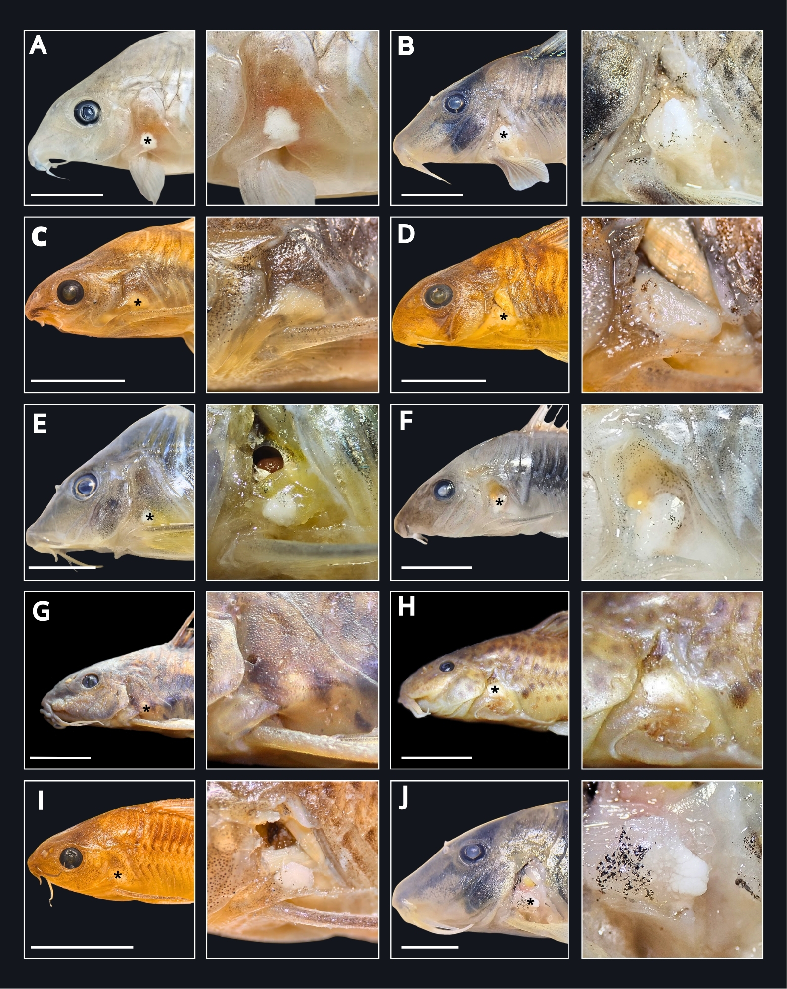
FIGURE 2 | Axillary glands in seven genera of Corydoradinae. Left column showing the position in relation to the head, with the axillary gland marked with an asterisk. Right column in a zoom showing the structure. A. Brochis sp.(non-catalogued); B. Corydoras simulatus (ICN 25509); C. Gastrodermus pygmeus (ICN 14814); D. Hoplisoma reynoldsi (ICN 29071); E. Brochis splendens (ICN 26511); F. Osteogaster aeneus (ICN 26507); G. Scleromystax macropterus (UFRGS 29647); H. Aspidoras fuscoguttatus (UFRGS 10522); I. Gastrodermus elegans (ICN 14252); J. Corydoras septentrionalis (ICN 26509). White scale bar represents 1 cm.

FIGURE 3 | Longitudinal section of the axillary gland of Brochis splendens (ICN 26511). A. General view of the gland and surrounding tissues. B. Connective tissue septa dividing the adjacent glandular lobules. C. Secretory cells of the axillary gland. D. Capsule and adjacent lobule of the axillary gland. Secretion (Sec), erythrocytes (Er), axillary gland cells (Agc), capsule (Cap). Black scale bar represents 100 µm.
Hoplisoma araguaiaense (Sands, 1990) displayed a distinctive pectoral spine morphology relative to other species of the genus. In this species, the spine was predominantly covered by an epidermis rich in club cells, along with an additional cell type lacking a visible nucleus and exhibiting a darker, occasionally granular cytoplasm.
No comparable glandular tissue was observed along the entire length of the pectoral spine in Corydoras, Aspidoras, Scleromystax, or Gastrodermus. In these genera, the spine was covered by an epidermis of variable thickness composed primarily of keratinocytes, goblet cells, and club cells, and occasionally containing eosinophilic granular secretory cells (Figs. 5E–H).
Within the clade comprising Aspidoras (A. albater Nijssen & Isbrücker, 1976) and Scleromystax (S. macropterus (Regan, 1913), S. virgulatus (Nijssen & Isbrücker, 1980)), the relatively thin pectoral spine was covered by an epidermis containing multiple layers of club cells measuring approximately 30–50 µm in diameter. These cells exhibited pale, homogeneous cytoplasm and centrally located nuclei and were present both along the spine and in its basal region (Figs. 5G–H). Club cells were more abundant in Aspidoras than in Scleromystax, although large aggregates were occasionally observed in the latter. In Gastrodermus elegans, the epidermis covering the pectoral spine was poorly developed and consisted predominantly of keratinocytes, with no evidence of specialized secretory cell types.

FIGURE 4 | Variation in the axillary gland architecture among Corydoradinae specimens. A. Axillary gland of Brochis delphax (ICN 26512), showing a lobular organization with granular secretion; Basophilic glandular cells release eosinophilic material. B. Axillary gland of Corydoras simulatus (ICN 25509) with uniformly granular secretion. C. Axillary gland of Hoplisoma reynoldsi (ICN 29071), characterized by glandular cells with a uniform appearance and pale pink cytoplasm in the periphery of the gland. D. Axillary gland of Hoplisoma metae (ICN 26508), displaying the coexistence of both secretory cell types. E. Axillary gland of Gastrodermus elegans (ICN 14252). F. Axillary gland of Corydoras pastazensis (ICN 10311). For each specimen, panels 1 and 2 represent magnifications of 100× and 400×, respectively. Uniform glandular cells (black asterisks), granular glandular cells (white asterisks), axillary glandular cells (Agc), secretion (Sec). Black scale represents 100 µm.
In Corydoras (C. pastazensis Weitzman, 1963, C. semiaquilus Weitzman, 1964, and C. simulatus Weitzman & Nijssen, 1970), the pectoral spine exhibited large serrations covered by a well-developed, highly secretory epidermis. This epidermis contained abundant eosinophilic granular secretory cells (20–50 µm in diameter) resembling those described for the axillary gland (Figs. 5E–F). The secretory epidermis extended along the entire length of the spine and included keratinocytes, blood vessels containing erythrocytes, and occasional club and goblet cells.

FIGURE 5 | Longitudinal histological sections of the pectoral spine showing differences between venom-bearing and non-venom-bearing genera of Corydoradinae. A. Brochis splendens (ICN 26511), showing the venom gland adjacent to the pectoral spine and covered by an epidermal layer. B. Hoplisoma metae (ICN 25479), with an epidermis containing granular secretory cells overlying the venom gland. C. Hoplisoma leucomelas (ICN 15840). D. Osteogaster melanotaenia. E. Corydoras simulatus (ICN 25509), in which no previously described venom gland is present; instead, a highly developed epidermis with abundant eosinophilic granular secretory cells occurs between the spine serrations. F. Corydoras semiaquilus (ICN 16865). G. Aspidoras albater (UFRGS 9906) displays a thin spine covered by a layer of club cells and keratinocytes. Stained with toluidine blue. H. Scleromystax virgulatus (UFRGS 11026). Stained with toluidine blue. For each specimen, panels 1 and 2 represent magnifications of 100× and 400×, respectively. Venom gland cells (Vgc), epidermis (Ep), pectoral spine (Ps), Club cells (Cc). Black scale bar represents 100 µm.
Discussion
Axillary gland. The axillary gland has been reported in several families of Siluriformes (Reed, 1907; Halstead et al., 1953; Whitear et al., 1991; Greven et al., 2006; Carvalho et al., 2021; Sodré et al., 2021; Egge, 2025), as well as in the toadfish family Batrachoididae (Reed, 1924; Maina et al., 1998; Wright, 2012). It is commonly described as a subcutaneous structure located beneath the posthumeral process of the cleithrum and typically associated with an axillary pore at the base of the pectoral spine. This pore is thought to serve as the external outlet for a multifunctional secretion released to the surrounding environment (Reed, 1907; Halstead et al., 1953; Birkhead, 1972; Whitear et al., 1991; Greven et al., 2006; Carvalho et al., 2021; Sodré et al., 2021). In some taxa, such as Hassar orestis (Steindachner, 1875), multiple pores are distributed throughout the axillary region, with each pore corresponding to a small individual gland (Reed, 1924). By contrast, in large-bodied catfishes such as Ictalurus punctatus (Rafinesque, 1818), axillary pores close early in ontogeny (Birkhead, 1972), whereas in heavily armored or naked species with reduced or loosely articulated spines, the pore is absent altogether (Reed, 1924). In Corydoradinae, the axillary gland is present in all examined genera and conforms to the described Siluriformes condition, being positioned beneath the cleithrum and associated with a single axillary pore.
Histologically, the axillary gland in Siluriformes has been described as sacciform (Wright, 2012) or pyriform (Reed, 1907; Halstead et al., 1953; Cameron, Endean, 1971; Carvalho et al., 2021), with reported sizes ranging from approximately 2.0 mm in Ariopsis felis (Linnaeus, 1766) (Halstead et al., 1953) to 5 mm in Noturus flavus Rafinesque, 1818and up to 20 mm in Cnidoglanis macrocephalus (Valenciennes, 1840) (Reed, 1907; Cameron, Endean, 1971). In Corydoradinae, axillary glands were predominantly round to oval, ranging from approximately 0.5 mm in smaller species to 2 mm in the largest examined specimen. These dimensions are consistent with those reported for similarly sized Siluriformes, such as Brachyrhamdia spp. (Carvalho et al., 2021). As in other catfishes, the glands were enclosed by a connective tissue capsule and often surrounded by adipose tissue. Connective tissue septa extended inward from the capsule, subdividing the gland into lobules or saccular units. Although a tubular organization has been previously described for one individual of Osteogaster aeneus (Gill, 1858)(Greven et al., 2006), our histological observations based on 51 individuals of Corydoradinae indicate a lobular architecture separated by connective septa (Fig. 3).
The cellular composition of the axillary gland consists primarily of secretory cells supported by a network of non-secretory cells. Secretory cells are the main constituent of the gland and are responsible for producing the glandular secretion. In Siluriformes, these cells are typically large and polygonal, ranging from 75 to 100 µm in diameter (Reed, 1924; Wright, 2012; Carvalho et al., 2021) and reaching up to 210 µm in A. felis (Halstead et al., 1953). They are characterized by large nuclei and a vacuolated cytoplasm that undergoes a progressive transformation from basophilic and frequently binucleated in immature stages to eosinophilic and anucleated in mature stages (Halstead et al., 1953; Cameron, Endean, 1971; Carvalho et al., 2021; Sodré et al., 2021; Egge, 2025). A similar gradient of cellular differentiation was observed in Corydoradinae. Immature cells display pale, occasionally binucleated cytoplasm, whereas more advanced stages were characterized by cells with granular cytoplasm and large nuclei that eventually disintegrated, releasing eosinophilic material into the glandular lobules. Different stages of cellular maturation were frequently observed within the same lobule, suggesting a continuous cycle of secretory cell differentiation and degeneration (Fig. 4).
Supporting cells were primarily located within the connective tissue septa, displaying elongated (filiform) nuclei and indistinct cytoplasm, forming a delicate scaffold surrounding the secretory cells (Cameron, Endean, 1971). In Corydoradinae, these septa were sparsely vascularized, in contrast to the highly vascularized condition reported for Acanthodoras spinosissimus (Eigenmann & Eigenmann, 1888)(Sodré et al., 2021), although some capillaries were occasionally observed. In addition, muscle fibers, adipose tissue, and immune cells were present around the axillary gland, consistent with previous descriptions in other siluriform species (Reed, 1907; Halstead et al., 1953; Cameron, Endean, 1971; Carvalho et al., 2021).
The glandular lumen of the axillary gland in Siluriformes has been described as extremely narrow or even absent, with no evidence of a well-defined duct for secretion transport. This condition suggests a holocrine mode of secretion, in which the secretory products are released when the secretory cells disintegrate (Cameron, Endean, 1971; Whitear et al., 1991; Carvalho et al., 2021). An exception has been reported for O. aeneus, in which a distinct lumen enabling glandular discharge was described (Greven et al., 2006). Our observations support the absence of a distinct lumen or duct in the axillary glands of Corydoradinae, further supporting a holocrine mechanism whereby secretory cells within each lobule disintegrate, releasing their contents and progressively filling the lobular cavity. The accumulated material was predominantly eosinophilic and occasionally formed reddish vesicles or a pink, granular secretion (Fig. 4). Under stress conditions, the secretion appeared transparent and contained minute white droplets, differing from the whitish, milky secretion reported for A. spinosissimus (Sodré et al., 2021). Personal observations under stress-induced conditions documented secretions with similar features in Corydoradinae species; nevertheless, future studies should expand protein quantification and identification analyses to elucidate the true nature of its function.
Axillary glands are present in all examined genera of Corydoradinae. However, we were unable to find evidence in the literature for the presence of axillary glands in Callichthyinae (Callichthys Scopoli, 1777, Dianema Cope, 1871, Hoplosternum Gill, 1858, Lepthoplosternum Reis, 1997, and Megalechis Reis, 1997)(Shimabukuro-Dias et al., 2004), the other subfamily of Callichthyidae. Nonetheless, axillary glands have been reported in multiple Siluriformes families (Egge, 2025), indicating that this structure is widely distributed across the catfish order. Despite its widespread distribution, the functional role of axillary gland secretions remains unresolved and has been the subject of long-standing debate (Egge, 2025). Proposed functions include antimicrobial activity, particularly against a group of aquatic bacteria (i.e., Vibrio spp.), which could reduce bacterial growth on the skin surface (Kiehl et al., 2006). Other authors have emphasized a defensive or crinotoxic function, whereby the secretion is released under stress to provoke aversive reactions in potential predators (Al-Hassan et al., 1987; Sodré et al., 2021; Egge, 2025). Additional hypotheses include roles in pheromonal communication, alarm signaling, or ionoregulation (Wright, 2012; Egge, 2025).
Recently, it has been suggested that axillary gland secretions exhibit similar toxicity levels between two species of Corydoradinae (H. metae and C. simulatus), based on a toxicity experiment that estimated lethality in shrimps (Phelps et al., 2026). These results indicate that toxicity remains a plausible function of this gland, particularly given that secretion release is commonly associated with stress responses. However, caution is warranted when interpreting these findings, as the natural predators of Corydoradinae are typically larger vertebrates (e.g., birds and larger fishes; Alexandrou et al., 2011), for which toxicity assessed using invertebrate models may not be directly comparable. Further studies focusing on the biochemical and proteomic composition of axillary gland secretions will be essential to elucidate their structural complexity and functional significance.
Pectoral-fin spine glands. In Siluriformes, venom delivery is generally mediated by the dorsal and pectoral spines. These spines possess an ossified core covered by a spine gland and integumentary sheath that releases its contents when the tissue is disrupted during interactions with predators (Halstead et al., 1953; Egge, 2025). The associated venom spine gland has been described as an aggregation of unicellular glandular cells adjacent to the bony spine, and it is thought to be derived from epidermal holocrine cells (Cameron, Endean, 1973). Numerous studies have documented the presence of venom glands associated with the dorsal and pectoral spines across Siluriformes (Reed, 1907, 1924; Halstead et al., 1953; Whitear et al., 1991; Carvalho et al., 2021; Egge, 2025).
This gland is typically described as a subcutaneous structure extending longitudinally along the pectoral spine, becoming progressively more abundant and better developed from the medial toward the distal portions. In A. felis, the gland is restricted to the distal two-thirds of the spine and gradually diminishes toward the base (Halstead et al., 1953), a pattern also observed in Noturus by Egge, Simons (2010), where the highest glandular density occurs in the distal third of the pectoral spine. Reed (1907) further noted that the gland is often associated with a swelling around the midsection of the spine and may occupy most of the space between the spine and the overlying skin. Similarly, Whitear et al. (1991), Bhimachar (1944), and Carvalho et al. (2021) reported that the gland lies immediately beneath the epidermis and directly adjacent to the bony spine in Heteropneustes fossilis Müller, 1840, and Brachyrhamdia spp.
In the Corydoradinae subfamily, in the genera Hoplisoma, Brochis, and Osteogaster, we observed a comparable distribution pattern, with glandular tissue most developed in the middle and distal portions of the pectoral spine and largely absent proximally. As reported for other Siluriformes, the gland in these taxa was situated directly against the bony spine and beneath the epidermal layer. Specific spatial arrangements of glandular tissue have been described in some species, such as A. felis, in which glandular cells occur along the anterolateral and posterolateral margins of the spine (Halstead et al., 1953), or along the posterior margin near the posterior serrae, as reported for Brachyrhamdia spp. (Carvalho et al., 2021). Wright (2012) documented extensive diversity in glandular arrangements across catfishes. In contrast, we observed glandular tissue along both the anterior and posterior margins of the first pectoral fin ray (the pectoral spine), without evidence of external swellings, longitudinal grooves, or depressions that might indicate the position of the gland, as reported for other taxa (Reed, 1907; Wright, 2012).
Venom gland cells associated with the pectoral spine are typically described as large (12–28 µm in A. felis) and formed by polygonal cells arranged in broad sheets or compact aggregates. Their cytoplasm is strongly acidophilic, likely reflecting a high protein content, and stains more intensely with eosin than surrounding club cells (Halstead et al., 1953; Whitear et al., 1991; Carvalho et al., 2021; Egge, 2025). Reed (1907) initially proposed that venom gland cells arise from the transformation of club cells; however, ultrastructural studies by Whitear et al. (1991) demonstrated that these represent distinct cell types. Although Egge, Simons (2010) noted morphological similarities between venom gland cells and club cells in Ictaluridae, they likewise considered them separate entities. In our material, when venom glands were present, club cells were restricted to the epidermis and exhibited a pale cytoplasm, whereas venom gland cells were located beneath the epidermis and displayed a markedly more eosinophilic cytoplasm.
Specifically, Wright (2009) reported contrasting results, documenting the presence of circumferential glandular cells associated with the pectoral spine in O. aeneus but absent in B. splendens. In contrast, our results support the presence of a shared venom gland morphology within a clade of Corydoradinae, in which the genera Osteogaster, Brochis, and Hoplisoma consistently exhibit this structure.
On the other hand, Corydoras exhibits a well-developed epidermis containing abundant keratinocytes, goblet cells, club cells, and conspicuous eosinophilic granular secretory cells, which closely resemble those of the axillary gland (Figs. 5E–F). These eosinophilic granular secretory cells were particularly abundant near the serrations and within the grooves separating the serrae. One possibility is that these cells correspond to the eosinophilic granular cells (EGCs) described by Mokhtar (2022), characterized by an eccentric nucleus and eosinophilic cytoplasmic granules, and generally associated with immune functions. However, EGCs have been mainly reported in the epidermis around the mouth and nasal fossae of fish. Alternatively, these cells may be homologous or functionally related to axillary gland secretory cells, potentially contributing to wound healing, local defense, or chemical signaling in the pectoral fin, an anatomical structure that is highly exposed and frequently prone to mechanical damage.
Gastrodermus exhibited a poorly developed epidermis over the pectoral spine, in some cases appearing completely absent, likely due to its small body size or to preservation artifacts. Aspidoras and Scleromystax form a clade characterized by similar epidermal morphology, dominated by club cells and keratinocytes, with no evidence of venom glands or eosinophilic granular secretory cells such as those observed in Corydoras. Wright (2009) reported the presence of circumferential glandular cells associated with the pectoral spine in Aspidoras albater; however, we were unable to confirm this condition in our material, where the epidermis of the spine instead contained numerous club cells (Fig. 5G).
The absence of venom gland morphology in the pectoral spine of Gastrodermus, Aspidoras,and Scleromystax supports the hypothesis that venom-bearing pectoral spine glands represent an evolutionary novelty within a clade of Corydoradinae, comprising Osteogaster, Brochis, and Hoplisoma (Fig. 6). Wright (2009) reported the presence of pectoral spine venom glands in other Callichthyidae, including Dianema longibarbis Cope, 1872, Hoplosternum littorale (Hancock, 1828), and Callichthys callichthys (Linnaeus, 1758), all members of the subfamily Callichthyinae. However, in the only species of this group illustrated by Wright (2009) (D. longibarbis, Fig. 2C), the venom gland cells are represented as a tissue composed of multiple round to polygonal cells with clear cytoplasm located within the epidermis. Based on their morphology and epidermal position, these cells more closely resemble epidermal club cells, which also exhibit clear cytoplasm and occur within the epidermis. To accept an alternative interpretation that venom-associated pectoral spine glands evolved multiple times within Callichthyidae, future analyses are needed to check if the glandular tissues reported by Wright (2009) in the pectoral fins of H. littorale and C. callichthys represent venom gland cells or epidermal club cells.

FIGURE 6 | Evolutionary distribution of axillary and pectoral spine glands in Corydoradinae. Simplified phylogeny is based on the molecular hypothesis proposed by Dias et al. (2024) and illustrates the presence of axillary glands across all genera of the subfamily, whereas the venom gland associated with the pectoral spine is restricted to a clade comprising Osteogaster, Brochis, and Hoplisoma. Because axillary glands also occur in other families of Siluriformes, their placement in the cladogram is intended for illustrative purposes only and does not represent a synapomorphy of Corydoradinae.
Implications in mimicry dynamics. Corydoradinae species have long been considered aposematic models due to their combination of an armored integument with a distinctive coloration pattern, the presence of sharp and lockable pectoral and dorsal spines, and the presence of toxic glands (Axenrot, Kullander, 2003; Alexandrou et al., 2011; Lima, Sazima, 2017). Together, these features are likely able to deter predators and constitute an effective evolutionary defense mechanism, making Corydoradinae an ideal system for investigating the ecology and evolutionary dynamics of freshwater mimicry (Ruxton et al., 2018). Importantly, our results indicate that while defensive traits are broadly distributed across the subfamily, they are not equivalent among genera, particularly in the presence of additional venom-associated pectoral spine glands, which may generate differences in defensive effectiveness and influence the type of mimicry involved.
The presence of venom-associated pectoral spine glands only in the clade comprising Brochis, Hoplisoma, and Osteogaster supports a single origin of these glands in the Corydoradinae (Fig. 6). Further, our histological analyses indicate that these glands are absent in Corydoras, Aspidoras, Scleromystax, and Gastrodermus. Interestingly, several species from both venom-bearing and non-venom-bearing lineages have been reported to participate in mimicry rings, often forming mixed-species shoals and exhibiting highly convergent color patterns. Alexandrou et al. (2011) identified 52 Corydoradinae species forming 24 putative Müllerian mimicry rings, with more than 90% of these rings composed of species from different genera that, nonetheless, share strikingly similar coloration. Classical Müllerian mimicry requires that all participating species are defended, contribute to predator deterrence, and share the costs associated with predator learning a common warning signal (Ruxton et al., 2018). Our results partially support this interpretation, as all examined Corydoradinae possess at least some combination of mechanical and glandular defenses. However, the additional presence of venom-associated pectoral spine glands in only some genera suggests that defensive effectiveness may not be equivalent among lineages.
The armored bodies covered with bony plates and odontodes, together with the ossified spines and their functional locking mechanism, constitute an effective mechanical defense that makes these fishes difficult to swallow, but does not completely prevent predation. For example, individuals of Corydoras have been reported among the dietary items of Hoplias malabaricus (Bloch, 1794), suggesting that these mechanical defenses can be overcome through predator behavior or by differences in predator-prey size (Loureiro, Hahn, 1996). This indicates that additional defensive mechanisms may further influence predator deterrence and raises important questions regarding the true nature of the proposed mimicry rings. A clear example is mimicry ring 11 described by Alexandrou et al. (2011), composed of C. simulatus and H. metae. Our histological evidence (Fig. 7) demonstrates that Corydoras lacks venom-associated pectoral spine glands, whereas Hoplisoma possesses them. Because both genera retain armored bodies, locking spines, and axillary glands, both are likely defended to some extent. However, the additional venom-associated pectoral spine glands in Hoplisoma suggest asymmetries in defensive effectiveness, indicating that this association may differ from a strictly Müllerian mimicry ring.
Previous studies have already suggested the occurrence of both types of mimicry within Corydoradinae and between Corydoradinae and other Siluriformes (Axenrot, Kullander, 2003; Lima, Sazima, 2017). A well-documented case involves Otocinclus mimulus Axenrot & Kullander, 2003 and Hoplisoma diphyes (Axenrot & Kullander, 2003) in the Paraguay basin, where the former is hypothesized to gain protection by mimicking the coloration of the latter. Hoplisoma diphyes possesses more effective antipredator traits, including a heavily armored body and lockable pectoral and dorsal spines, whereas O. mimulus lacks these defenses (Axenrot, Kullander, 2003). A similar pattern was suggested to occur within Corydoradinae itself. Lima, Sazima (2017) proposed that some species of Corydoras such as C. desana Lima & Sazima, 2017, act as Batesian mimics of species of Hoplisoma, such as H. tukano (Britto & Lima, 2003). In this case, the unpalatable trait of H. tukano is attributed to its sharper, more penetrating pectoral spine, which would release the venom more efficiently in the predator, in contrast to the highly serrated but apparently less pungent spine of C. desana. In both scenarios, the more defended species is more frequent than the mimic, allowing the warning signal to remain reliably within the predator community, one of the key conditions for Batesian mimicry. However, because both species retain armored bodies, lockable fin spines, and likely defensive axillary gland secretions, both could be considered defended to some extent. Nevertheless, defensive venoms that result in tissue damage or neurological dysfunction may be required to elicit greater aversion than venoms that cause pain alone (Niermann et al., 2020).
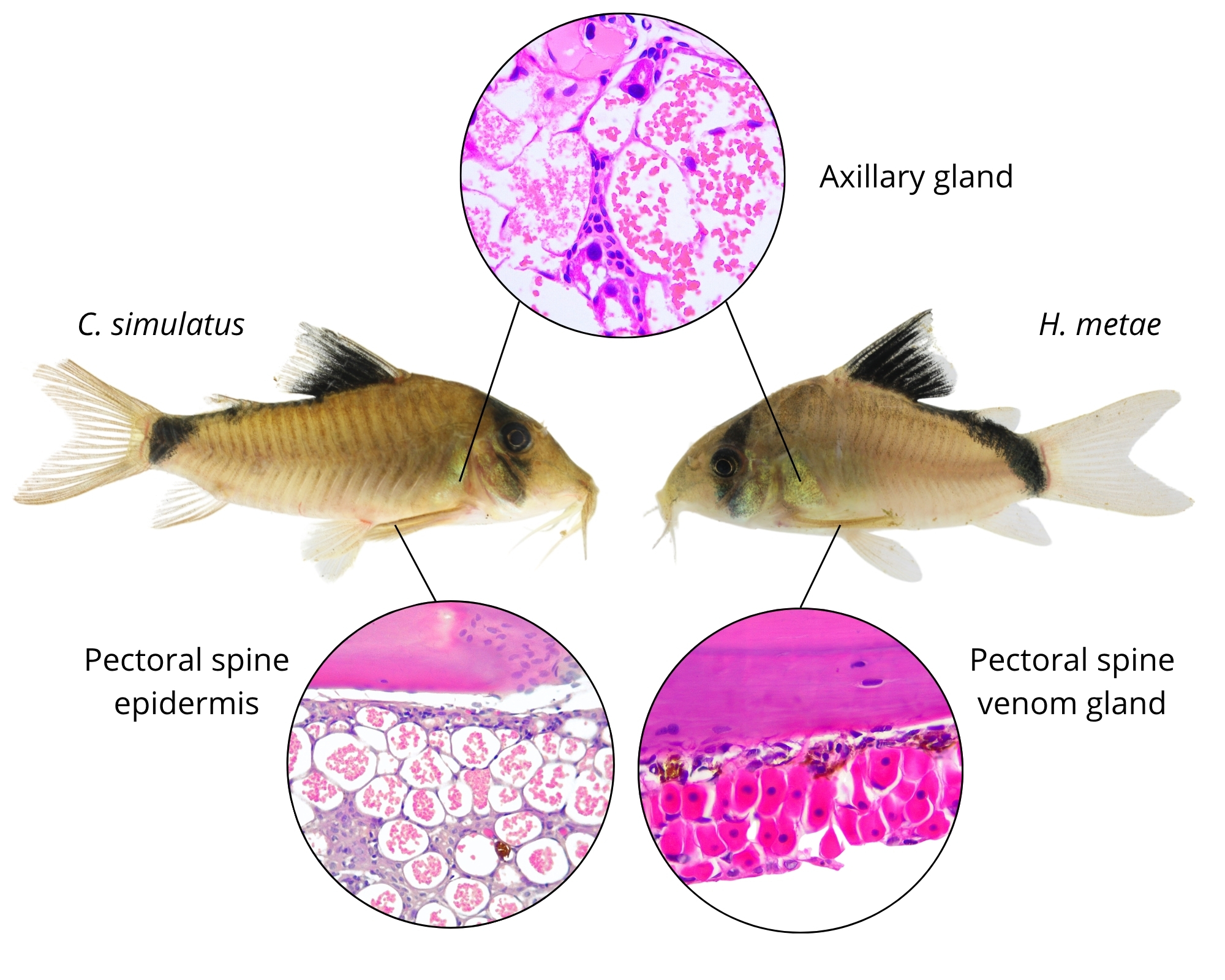
FIGURE 7 | Mimicry ring involving Corydoras simulatus and the venomous Hoplisoma metae co-occurring in the Meta River basin. Although both possess axillary glands of similar morphology, they differ in the presence of venom spine glands.
Importantly, our histological analyses reveal clear intergeneric differences in the presence of venom-associated pectoral spine glands, indicating that some genera possess more effective defensive traits than others. Building on these considerations, our results support the existence of mixed mimicry systems within Corydoradinae, in which unequally defended species coexist sympatrically and share similar aposematic color patterns. In this context, the concept of quasi-Batesian mimicry may better explain these interactions. Under this framework, all participating species are defended to some extent, but one species possesses stronger antipredator defenses than the other. As proposed by Speed (1993), quasi-Batesian mimicry occurs when unequally defended species converge on a shared warning signal: both contribute to predator learning through a Müllerian component, while the less defended species may also benefit disproportionately from resembling the more strongly defended model, introducing a Batesian component to the interaction (Speed, 1993; Ruxton et al., 2018).
Consequently, in assemblages with three mimetic species, where two strongly defended coexist with a less defended species exhibiting similar coloration, an interesting case of mixed mimicry ring may arise, in which equally defended taxa participate in Müllerian mimicry while non-equally defended taxa function as quasi-Batesian mimics [e.g., B. imitator (Nijssen & Isbrücker, 1983), H. adolfoi (Burgess, 1982), and G. nijsseni (Sands, 1989) in mimicry ring 12 of Alexandrou et al., 2011]. However, quasi-Batesian interactions may shift toward classical Batesian mimicry when predators experience nutritional stress and the less defended species becomes profitable to consume. Under these conditions, the less defended species may function temporarily as a true Batesian mimic, despite retaining some defensive traits (Ruxton et al., 2018).
Incorporating the concept of quasi-Batesian mimicry into the Corydoradinae system suggests that mimicry in this subfamily may represent a continuum between Müllerian and Batesian interactions, raising new questions about the ecological and evolutionary mechanisms underlying these associations. Future studies should evaluate the relative abundance of co-mimetic shoals, the occurrence of polymorphic colour patterns within local assemblages, and predator responses under different nutritional states, as these factors may strongly influence the stability and selective dynamics of mimicry systems.
In conclusion, our study demonstrates that axillary glands are a conserved and structurally consistent feature across Corydoradinae, exhibiting a lobular, holocrine organization comparable to that of other Siluriformes, although their precise functional role remains unresolved and likely multifaceted. In contrast, venom-associated pectoral spine glands show a more restricted phylogenetic distribution, occurring only within a specific clade (Brochis, Hoplisoma, and Osteogaster), which supports a single evolutionary origin of this defensive trait within the subfamily. This heterogeneity in defensive mechanisms has important ecological and evolutionary implications, particularly for mimicry systems, as it challenges the assumption that all Corydoradinae species involved in mimicry rings are equally defended. Instead, our findings support the presence of Müllerian, quasi-Batesian, and Batesian mimicry components within the group, depending on asymmetries in defensive effectiveness among participating taxa, particularly the presence or absence of venom-associated pectoral spine glands.
Acknowledgments
We are especially grateful to Yecid Rojas for his assistance during fieldwork, and to the ornamental fishers Tobias and Josué OCampo from the communities of Acacías and Puerto Lleras, Meta, Colombia, for their kindness and invaluable help during specimen collection. We also thank Tiago P. Carvalho, curator of the ichthyological collection at the Instituto de Ciencias Naturales, Universidad Nacional de Colombia, for his guidance and collaboration in species identification, and Mery Martínez García and Julia Giora for their valuable recommendations regarding histological processing and analysis.
References
Alexandrou MA, Oliveira C, Maillard M, McGill RAR, Newton J, Creer S et al. Competition and phylogeny determine community structure in Müllerian co-mimics. Nature. 2011; 469:84–88. https://doi.org/10.1038/nature09660
Al-Hassan JM, Thomson M, Ali M, Criddle RS. Toxic and pharmacologically active secretions from the Arabian Gulf catfish (Arius thalassinus, Ruppell). J Toxicol Toxin Rev. 1987; 6(1):1–43. https://doi.org/10.3109/15569548709053860
Alonso F, Terán GE, Aguilera G, Montes MM, Serra Alanís WS, Calviño P et al. Integrative phylogeny of Corydoradinae (Siluriformes: Callichthyidae) with an emphasis on northwestern La Plata species, including descriptions of a new genus and species. Zool Anz. 2025; 314:10–23. https://doi.org/10.1016/j.jcz.2024.11.006
Axenrot TE, Kullander SO. Corydoras diphyes (Siluriformes: Callichthyidae) and Otocinclus mimulus (Siluriformes: Loricariidae), two new species of catfishes from Paraguay, a case of mimetic association. Ichthyol Explor Freshw. 2003; 14(3):249–72.
Bhimachar BS. Poison glands in the pectoral spines of two catfishes – Heteropneustes fossilis (Bloch) and Plotosus arab (Forsskål), with remarks on the nature of their venoms. Proc Indian Natl Sci Acad B Biol Sci. 1944; 19:65–70.
Birkhead WS. Toxicity of stings of Ariid and Ictalurid catfishes. Copeia. 1972; 1972(4):790–807. https://doi.org/10.2307/1442737
Britto MR. Phylogeny of the subfamily Corydoradinae Hoedeman, 1952 (Siluriformes: Callichthyidae), with a definition of its genera. PNAS. 2003; 153(1):119–54. https://doi.org/10.1635/0097-3157(2003)153[0119:POTSCH]2.0.CO;2
Cameron AM, Endean R. The axillary glands of the plotosid catfish Cnidoglanis macrocephalus. Toxicon. 1971; 9(4):345–52. https://doi.org/10.1016/0041-0101(71)90132-2
Cameron AM, Endean R. Epidermal secretions and the evolution of venom glands in fishes. Toxicon. 1973; 11(5):401–410. https://doi.org/10.1016/0041-0101(73)90115-3
Carvalho TI, Klaczko J, Slobodian V. Pectoral-fin glands and delivery apparatus in the catfish genus Brachyrhamdia Myers, 1927 (Siluriformes: Heptapteridae). Pap Avulsos Zool. 2021; 61:e20216174. https://doi.org/10.11606/1807-0205/2021.61.74
Casewell NR, Wüster W, Vonk FJ, Harrison RA, Fry BG. Complex cocktails: the evolutionary novelty of venoms. Trends Ecol Evol. 2012; 28(4):219–29. https://doi.org/10.1016/j.tree.2012.10.020
Dias AC, Tencatt LFC, Roxo FF, Silva GSC, Santos SA, Britto MR et al. Phylogenomic analyses in the complex Neotropical subfamily Corydoradinae (Siluriformes: Callichthyidae) with a new classification based on morphological and molecular data. Zool J Linn Soc. 2024; 203(3):zlae053. https://doi.org/10.1093/zoolinnean/zlae053
Egge JJ. Siluriform venom system. In: Arratia G, Reis RE, editors. Catfishes, a highly diversified group. Volume 1: their outstanding biology. 1st ed. Boca Raton: CRC Press; 2025. p.160–81. https://doi.org/10.1201/9781003361336
Egge JJD, Simons AM. Evolution of venom delivery structures in madtom catfishes (Siluriformes: Ictaluridae). Biol J Linn Soc. 2010; 102(1):115–29. https://doi.org/10.1111/j.1095-8312.2010.01578.x
Greven H, Flasbeck T, Passia D. Axillary glands in the armoured catfish Corydoras aeneus (Callichthyidae, Siluriformes). Verh Ges Ichthyol. 2006; 5:65–69.
Halstead BW. Poisonous and venomous marine animals of the world. 2nd rev. ed. Princeton: The Darwin Press, Inc.; 1988.
Halstead BW, Kuninobu LS, Hebard HG. Catfish stings and the venom apparatus of the Mexican catfish, Galeichthys felis. Trans Am Microsc Soc. 1953; 72(4):297–314. https://doi.org/10.2307/3223475
Harris RJ, Jenner RA. Evolutionary ecology of fish venom: adaptations and consequences of evolving a venom system. Toxins. 2019; 11:60. https://doi.org/10.3390/toxins11020060
Hildebrandt J, Teuscher E, Lindequist U. Introduction to animal toxins. In: Hildebrandt J, Teuscher E, Lindequist U, editors. Natural poisons and venoms: animal toxins. Vol. 4. Berlin: De Gruyter; 2023. p.1–17.
Kiehl E, Rieger C, Greven H. Axillary gland secretions contribute to the stress-induced discharge of a bactericidal substance in Corydoras sterbai (Callichthyidae, Siluriformes). Verh Ges Ichthyol. 2006; 5:111–15.
Lima FCT, Sazima I. Corydoras desana, a new plated catfish from the upper rio Negro, Brazil, with comments on mimicry within the Corydoradinae (Ostariophysi: Siluriformes: Callichthyidae). Aqua. 2017; 23(1):19–23.
Loureiro VE, Hahn NS. Dieta e atividade alimentar da traíra, Hoplias malabaricus (Bloch, 1794) (Osteichthyes, Erythrinidae), nos primeiros anos de formação do reservatório de Segredo – PR. Acta Limnol Brasil. 1996; 8:195–205.
Maina JN, Wood CM, Hogstrand C, Hopkins TE, Luo YH, Gibbs PDL et al. Structure and function of the axillary organ of the Gulf toadfish, Opsanus beta (Goode and Bean). Comp Biochem Physiol A Mol Integr Physiol. 1998; 119(1):17–26. https://doi.org/10.1016/S1095-6433(97)00410-8
Mokhtar DM. Skin and associated sense structures. In: Mokhtar DM, editors. Fish histology: from cells to organs. 2nd ed. Palm Bay: Apple Academic Press; 2022. p.99–136.
Ndandala CB, Mustapha UF, Wang Y, Assan D, Zhao G, Huang C et al. The perspective of fish venom: an overview of the physiology, evolution, molecular and genetics. Front Mar Sci. 2023; 10:1085669. https://doi.org/10.3389/fmars.2023.1085669
Nelsen DR, Nisani Z, Cooper AM, Fox GA, Gren ECK, Corbit AG et al. Poisons, toxungens, and venoms: redefining and classifying toxic biological secretions and the organisms that employ them. Biol Rev. 2014; 89(2):450–65. https://doi.org/10.1111/brv.12062
Niermann CN, Tate TG, Suto AL, Barajas R, White HA, Guswiler OD et al. Defensive venoms: is pain sufficient for predator deterrence? Toxins. 2020; 12(4):260. https://doi.org/10.3390/toxins12040260
Perrière C, Goudey-Perrière F. Poisonous catfishes: venom apparatus, acanthotoxins, crinotoxins, and other skin secretions. In: Arratia G, Kapoor BG, Chardon M, Diogo R, editors. Catfishes: Volume 1. Enfield: Science Publishers; 2003. p.291–314.
Phelps E, Bell E, Immler S, Taylor MI. Axillary gland gene expression and potency reveal the nature of mimetic relationships in Corydoradinae catfishes. bioRxiv [Preprint]. 2026. https://doi.org/10.64898/2026.01.27.701968
Reed HD. The poison glands of Noturus and Schilbeodes. Am Nat. 1907; 41:53–566. https://www.jstor.org/stable/2455004
Reed HD. The morphology of the dermal glands in nematognathous fishes. Z Morphol Anthropol. 1924; 24:227–64. http://www.jstor.org/stable/25748873
Ross MH, Pawlina W. Techniques. In: Ross MH, Pawlina, editors. Histology: a text and atlas: with correlated cell and molecular biology. 8th ed. Philadelphia: Wolters Kluwer; 2018. p.1–27.
Ruxton GD, Allen WL, Sherratt TN, Speed MP. Avoiding the attack: the evolutionary ecology of crypsis, aposematism, and mimicry. 2nd ed. Oxford: Oxford University Press; 2018.
Sodré JG, Colaço-Fernandes TR, Rapp Py-Daniel LH, Birindelli JLO, Zuanon J. Putative poison gland in the thorny catfish Acanthodoras spinosissimus (Siluriformes: Doradidae). Acta Amazon. 2021; 51(3):250–54. https://doi.org/10.1590/1809-4392202101350
Shimabukuro-Dias CK, Oliveira C, Reis RE, Foresti F. Molecular phylogeny of the armored catfish family Callichthyidae (Ostariophysi, Siluriformes). Mol Phylogenet Evol. 2004; 32(1):152–63. https://doi.org/10.1016/j.ympev.2003.11.010
Smith WL, Stern JH, Girard MG, Davis PM. Evolution of venomous cartilaginous and ray-finned fishes. Integr Comp Biol. 2016; 56(5):950–61. https://doi.org/10.1093/icb/icw070
Smith WL, Wheeler WC. Venom evolution widespread in fishes: a phylogenetic road map for the bioprospecting of piscine venoms. J Hered. 2006; 97(3):206–17. https://doi.org/10.1093/jhered/esj034
Speed MP. Mullerian mimicry and the psychology of predation. Anim Behav. 1993, 45:571–80.
Whitear M, Zaccone G, Ricca MB, Lo Cascio P. Fine structure and histochemistry of the venom gland in the Indian stinging catfish (Heteropneustes fossilis). J Zool. 1991; 224(3):479–89. https://doi.org/10.1111/j.1469-7998.1991.tb06037.x
Wright JJ. Diversity, phylogenetic distribution, and origins of venomous catfishes. BMC Evol Biol. 2009; 9:282. https://doi.org/10.1186/1471-2148-9-282
Wright JJ. The evolutionary ecology of venomous catfishes, with a focus on members of the North American family Ictaluridae (Teleostei: Siluriformes). [PhD Thesis]. Ann Arbor: University of Michigan; 2012. Available from: https://hdl.handle.net/2027.42/93934
Wright JJ. Evolutionary history of venom glands in the Siluriformes. In: Gopalakrishnakone P, Malhotra A, editors. Evolution of venomous animals and their toxins. Dordrecht: Springer; 2015. p.1–19. https://doi.org/10.1007/978-94-007-6727-0_9-1
Ziegman R, Alewood P. Bioactive components in fish venoms. Toxins. 2015; 7(5):1497–531. https://doi.org/10.3390/toxins7051497
Authors

Camilo R. Jiménez-Vergara1 , Andréa Tonolli Thomaz1, Angélica J. Garzón-Restrepo2, Luiz R. Malabarba3 and Mario Monroy1
[1] Departamento de Biología, Facultad de Ciencias, Universidad Nacional de Colombia, Sede Bogotá, Carrera 45, No 26-85, 111321, Bogotá D.C, Colombia. (CRJV) crjimenezve@unal.edu.co (corresponding author), (ATT) atonolli@unal.edu.co, (MM) mamonroyl@unal.edu.co.
[2] Laboratorio de Virología y Biología Molecular, Facultad de Medicina Veterinaria y de Zootecnia, Universidad Nacional de Colombia, Sede Bogotá, Carrera 45, No 26-85, 111321, Bogotá D.C, Colombia. (AJGR) ajgarzonr@unal.edu.co.
[3] Programa de Pós-Graduação em Biologia Animal, Universidade Federal do Rio Grande do Sul, Av. Bento Gonçalves, 9500, 91501 970, Porto Alegre, RS, Brazil. (LRM) malabarb@ufrgs.br.
Authors’ Contribution 

Camilo R. Jiménez-Vergara: Conceptualization, Data curation, Formal analysis, Investigation, Methodology, Visualization, Writing-original draft, Writing-review and editing.
Andréa Tonolli Thomaz: Conceptualization, Formal analysis, Investigation, Methodology, Supervision, Writing-review and editing.
Mario Monroy: Conceptualization, Formal analysis, Investigation, Methodology, Supervision, Funding acquisition, Writing-review and editing.
Luiz R. Malabarba: Resources, Investigation, Writing-review and editing.
Angélica J. Garzón-Restrepo: Investigation, Methodology, Writing-review and editing.
Ethical Statement
The experimental procedures involving animals were approved by the Ethics Committee for Animal Experimentation (CEPI) of the Universidad Nacional de Colombia (Approval No 21688). All fish were euthanized in accordance with the ethical standards and procedures established under Colombian legislation. Specimens collected during fieldwork were obtained under the institutional framework collection permit granted by the Autoridad Nacional de Licencias Ambientales (ANLA) through Resolution No 255 de 2014, in accordance with Colombian environmental regulations.
Competing Interests
The author declares no competing interests.
Data availability statement
All data supporting the findings of this study are available within the article and its supplementary material. Histological photographs underlying the analyses are available from the corresponding author upon request. Examined specimens are deposited in institutional ichthyological collections as indicated in the text.
AI statement
Grammarly Premium (AI-assisted writing software) was used exclusively for English language editing and grammar correction. The tool was employed only to improve language clarity and readability. It was not used for study design, data analysis, interpretation of results, figure preparation, or the generation of scientific content. All scientific ideas, analyses, and conclusions presented in this manuscript are the original work of the authors.
Funding
This research was supported by the “Convocatoria conjunta entre la Universidad del Rosario y la Universidad Nacional de Colombia sede Bogotá para proyectos de investigación, innovación y creación – 2023” (Modalidad 1: Formativa y de iniciación a la investigación, innovación o creación).
Supplementary Material
Supplementary material SUP
Peer Review
How to cite this article
Jiménez-Vergara CR, Thomaz AT, Garzón-Restrepo AJ, Malabarba LR, Monroy M. Morphological diversification of defensive glands in Corydoradinae catfishes indicates a mixed Müllerian-Batesian mimicry system. Neotrop Ichthyol. 2026; 24(4):e260051. https://doi.org/10.1590/1982-0224-2026-0051
Copyright
This is an open access article under the terms of the Creative Commons Attribution License, which permits use, distribution and reproduction in any medium, provided the original work is properly cited.
Distributed under
Creative Commons CC-BY 4.0

© 2025 The Authors.
Diversity and Distributions Published by SBI
 Accepted August 14, 2026
Submitted April 7, 2026
Epub October 19, 2026