
 Sérgio João Sartor Neto1, Ligia Carla Balini2, Lucas Pietro Ferrari Gianini3, Cláudio Henrique Zawadzki3,4, Fernanda Errero Porto5, Luciana Andreia Borin-Carvalho1,2, Alessandra Valéria de Oliveira1,4,6, Laura Ivana Ramos6 and Carlos Alexandre Fernandes1,3,4
PDF: Download Here | Supplementary: Sup | Cite this article
Associate Editor: Claudio Oliveira
Section Editor: Izeni Farias
Editor-in-chief: José Birindelli
Abstract
A escassez de dados citogenéticos e genéticos sobre Rineloricaria langei destaca a necessidade de investigações abrangentes. Este estudo fornece a primeira caracterização citogenética da espécie e apresenta novos dados genéticos baseados em uma população amostrada de sua localidade-tipo. As análises citogenéticas revelaram dois cariomorfos com números diploides de 2n = 72, o maior relatado para o gênero, e 2n = 70. Apesar das diferenças na composição do cariótipo, ambos compartilham características estruturais semelhantes, incluindo heterocromatina predominantemente pericentromérica, um único sítio de rDNA 18S e múltiplos sítios de rDNA 5S, conforme detectado por hibridização fluorescente in situ (FISH). As análises moleculares identificaram três haplótipos (H1–H3) de R. langei, com uma divergência máxima de 0,4%. A inclusão de sequências públicas de COI aumentou o total para 18 haplótipos e revelou distâncias interespecíficas superiores a 4%, identificando R. zawadzkii como a espécie irmã. As análises de delimitação de espécies (ASAP, PTP e GMYC) recuperaram de 10 a 12 OTUs para o gênero, mas delimitaram consistentemente uma única OTU para R. langei. Esses achados revelam polimorfismo cromossômico intrapopulacional associado à baixa divergência genética mitocondrial e reforçam a importância de abordagens integrativas para a compreensão da variabilidade genética e estrutural em R. langei.
Palavras-chave: Código de barras de DNA, Diversidade genética, DNA ribossomal, Espécies crípticas, Evolução cariotípica.
Introduction
The order Siluriformes encompasses a broad array of freshwater fishes, including representatives of the family Loricariidae, which is recognized as one of the most speciose and diverse groups within the Neotropical region. Currently, Loricariidae is subdivided into six subfamilies, of which Loricariinae is the second largest, comprising 277 valid species (Fricke et al., 2025a). Members of the Loricariinae, commonly referred to as “guitar catfish” or “slipper catfish”, are distinguished by their dorsoventrally flattened bodies, absence of an adipose fin, and an elongated, depressed caudal peduncle (Rapp Py-Daniel, 1997). Within this subfamily, the genus Rineloricaria Bleeker, 1862, includes 77 valid species (Fricke et al., 2025b), with a distribution extending from Central America to southern Brazil and northern Argentina (Vera-Acaraz et al., 2012). Moreover, this genus is notable for its exceptional diversity and pronounced taxonomic complexity within the subfamily, a circumstance that is further complicated by unresolved questions concerning its geographic origins (Fichberg, 2008).
Recent advancements in cytogenetic and molecular methodologies have substantially enhanced the identification of poorly defined or cryptic species (Rosso et al., 2012). Karyotype analysis, for instance, facilitates the detection of intraspecific variation, including changes in diploid number, karyotypic formula, heterochromatin distribution patterns, and the localization of repetitive DNA sequences, parameters that are fundamental for precise species identification (Angulo, Icochea, 2010). In addition, molecular approaches such as DNA barcoding, which relies on the sequencing of the cytochrome c oxidase, subunit I (COI) gene, have demonstrated considerable efficacy in species delimitation and taxonomic identification, particularly within highly complex groups such as fishes (Morais-Silva et al., 2018; Soria et al., 2018). This technique is broadly applicable among eukaryotes, as it permits sequencing across diverse taxa using a limited set of primers (Hebert et al., 2003; Pereira et al., 2013).
Due to the considerable diversity and taxonomic complexity of the genus Rineloricaria, as well as the limitations of traditional morphological analyses in accurately delineating its species, the integration of multiple methodological approaches is essential for a comprehensive understanding of its diversity. Cytogenetic and molecular studies serve as valuable complementary tools by enabling the detection of chromosomal polymorphisms, the identification of cryptic species, and the inference of evolutionary processes (Morais et al., 2024). Cytogenetic investigations of the Rineloricaria have predominantly focused on the chromosomal characterization of 18 species distributed across various river basins in southern and south-eastern Brazil (Sassi et al., 2024), which constitutes approximately 23% of the currently recognized species diversity within the genus. Despite the limited cytogenetic data available for Rineloricaria, considerable inter- and intraspecific chromosomal variability has been documented. The diploid chromosome number (2n) has been observed to range from ♂33/♀34 in R. teffeana (Steindachner, 1879) (Marajó et al., 2023) up to 70 chromosomes in R. lima (Kner, 1853) (Rosa et al., 2012), Rineloricaria n. sp. (sensu Alves et al., 2003), R. longicauda Reis, 1983, and R. quadrensis Reis, 1983 (Venturelli et al., 2021). At the intraspecific level, pronounced structural polymorphisms have been reported, such as in R. latirostris (Boulenger 1900), which displays a chromosome number ranging from 46 to 48 (Glugoski et al., 2018; 2022), in R. lima, ranging from 66 to 70 chromosomes (Rosa et al., 2012), and in R. lanceolata (Günther, 1868), ranging from 45 to 48 chromosomes (Porto et al., 2014).
Within the 77% of Rineloricaria species that remain cytogenetically uncharacterized, Rineloricaria langei Ingenito, Ghazzi, Duboc & Abilhoa, 2008, is found in the Iraí River, a tributary of the upper Iguaçu River headwaters situated in the metropolitan region of Curitiba, Paraná State, Brazil. This species inhabits small streams with light to moderate water flow, typically over substrates composed of sand, rocks, and decomposing plant material (Ingenito et al., 2008). Morphologically, R. langei exhibits a slender body and bears resemblance to R. quadrensis, a species distributed in the coastal rivers of Rio Grande do Sul State; however, the two species can be distinguished based on morphometric characteristics (Ingenito et al., 2008).
The scarcity of cytogenetic and genetic data regarding R. langei underscores the necessity for more comprehensive investigations. Accordingly, this study sought to provide the first cytogenetic characterization of this species and to report genetic data that remain scarce in the existing literature, based on a population of R. langei sampled from its type locality in the Iraí River (upper Iguaçu River basin). Classical and molecular cytogenetic methodologies, along with analyses of the mitochondrial COI gene, were employed to evaluate intraspecific variation and provide data relevant to the taxonomic delimitation and conservation of the species.
Material and methods
Study area and sampling. A total of twenty individuals of R. langei (Siluriformes, Loricariidae), comprising twelve females and eight males, were collected from the Iraí River in the upper Iguaçu River basin (Fig. 1), which constitutes the type locality of the species. This site is situated in the municipality of Piraquara, Paraná State, Brazil, 25°28’49.2”S 49°06’12.8”W. Voucher specimens were deposited in the fish collection of the Núcleo de Pesquisas em Limnologia, Ictiologia e Aquicultura (NUP), Universidade Estadual de Maringá (UEM), Brazil, as R. langei (NUP 26569) (Fig. 1).

FIGURE 1| Location of Iraí River in the Iguaçu River basin where Rineloricaria langei (photo) individuals were captured. The red circle indicates the sampling spot.
Cytogenetic analysis. Metaphase chromosomes were obtained from anterior kidney cells using the air-drying technique (Bertollo et al., 2015). The nucleolus organizer regions (NORs) were detected employing silver nitrate staining (Howell, Black, 1980). Constitutive heterochromatin was determined following the C-banding technique (Sumner, 1972) and stained with propidium iodide (Lui et al., 2012).
At least 30 metaphases were analyzed for each individual, and those with better chromosome morphology were used for the karyotype analysis. The chromosomes were classified as metacentric (m), submetacentric (sm), subtelocentric (st), and acrocentric (a) according to Levan et al. (1964). The fundamental number (FN) was calculated according to the chromosomal arm numbers (the chromosomes m, sm, and st were considered to contain two arms, and the a chromosomes with one arm).
The location of the rDNA sites in the chromosomes was performed by fluorescence in situ hybridization (FISH) with modifications (Margarido, Moreira-Filho, 2008; Pinkel et al., 1986) using probes from the genome of Megaleporinus elongatus (Valenciennes, 1850) (Martins, Galetti Jr., 1999) for 5S rDNA sites and Prochilodus argenteus Spix & Agassiz, 1829 (Hatanaka, Galetti, 2004) for 18S rDNA sites. The probes were labeled through nick translation with digoxigenin-11-dUTP (5S rDNA) and biotin-16-dUTP (18S rDNA) (Roche Diagnostics, Mannheim, Germany). Detection and amplification of the hybridization signal were carried out using avidin-FITC and anti-avidin biotin (Sigma-Aldrich, St. Louis, Missouri, United States) for probes labeled with biotin and anti-digoxigenin rhodamine (Roche Diagnostics, Mannheim, Germany) for probes labeled with digoxigenin. Chromosomes were counterstained with 4’-6-diamin-2-phenylindole (DAPI, 50 μg ml−1, Sigma-Aldrich, St. Louis, Missouri, United States).
Conventional and fluorescence chromosome preparations were analyzed under a BX 51 epifluorescence microscope (Olympus America, Inc.). The images were captured using the DP controller (Media Cybernetics) software and composed in Adobe Photoshop CS6.
Molecular analysis. Genomic DNA was extracted from liver tissue using the Promega Wizard® Genomic DNA Purification Kit according to the manufacturer’s protocol. DNA concentration was estimated for each sample utilizing a NanoDrop™ Lite spectrophotometer (Thermo Scientific).
The cytochrome c oxidase subunit I (COI) gene was subsequently amplified using the FishF121 (Ward et al., 2005) and FR1d (Ivanova et al., 2007) primer pair. Polymerase chain reactions (PCR) were carried out in 0.2 mL microtubes in a final volume of 25 µL, containing approximately 10 ng of template DNA, 1× Tris–KCl buffer (10 mM Tris-HCl, pH 8.5, 50 mM KCl), 1.5 mM MgCl₂, 0.2 mM of each dNTP, 0.5 µM of each primer, 1.25 U of Taq DNA polymerase, and Milli-Q water to complete the final volume. The amplification protocol comprised 35 cycles: denaturation at 94ºC for 30 s, annealing at 50ºC for 1 min 30 s, and extension at 72ºC for 1 min, followed by a final extension at 72ºC.
Following amplification, PCR products were resolved on a 1% agarose gel, and fragment sizes were estimated using a 100 bp DNA ladder as a molecular size standard. PCR products were purified according to the protocol described by Rosenthal et al. (1993). Sequencing reactions were conducted using the BigDye Terminator kit, and sequencing was carried out to a private company using an AB 3500 automated sequencer.
For data analysis, sequence editing and alignment were conducted using BioEdit 7 and MEGA 7, respectively (Hall, 1999; Kumar et al., 2016), with Clustal W employed as the alignment algorithm (Thompson et al., 1994). Sequence identity percentages were determined using the BLASTn tool by comparison with reference sequences available in GenBank.
Additional COI sequences of Rineloricaria available in public databases were incorporated, with five sequences per species selected, prioritizing taxa genetically closer to R. langei. Sequences that negatively affected the final alignment length were replaced. Intraspecific and interspecific genetic distance (p-value) were calculated. The optimal evolutionary model was identified using MEGA 7, and a gene tree was constructed using the maximum likelihood method with 1,000 bootstrap replicates. Intrapopulation polymorphism was assessed using DNA Sequence Polymorphism (DnaSP v. 6) (Rozas et al., 2017).
Four species delimitation methods were employed: Assemble Species by Automatic Partitioning (ASAP) (Puillandre et al., 2021), the Poisson Tree Processes model (PTP) (Zhang et al., 2013), and the General Mixed Yule Coalescent approach (GMYC) (Fujisawa, Barraclough, 2013). For the GMYC delimitation analysis, an ultrametric phylogenetic tree was inferred under a Bayesian framework using a strict molecular clock and a constant-size coalescent model, on an arbitrary timescale, in BEAST v. 1.8.4 (Drummond et al., 2012), with input files generated in BEAUti. An initial random tree was used for the MCMC searches, which were run for 10 million generations, sampling one tree every 1,000 generations. Chain convergence and sampling adequacy were assessed in Tracer v. 1.7.1 (Rambaut et al., 2018), ensuring effective sample size (ESS) values above 200 for all parameters. Trees sampled prior to stationarity were discarded as burn-in (10%) in TreeAnnotator v. 1.8.4, and a maximum clade credibility tree was generated and used in subsequent analyses. The ASAP analysis was performed in a Python environment using the script delimitacao.py, based on the distance matrices generated for the dataset, and partitions were selected according to ASAP score values. The PTP analysis was conducted on the web server (https://species.h-its.org/ptp/). GMYC species delimitation was performed on the GMYC web server (https://species.h-its.org/gmyc/) using the single-threshold model, with all other parameters set to default.
Access to the genetic heritage of the specimens was authorized by the Sistema Nacional de Gestão do Patrimônio Genético (SISGEN; registration A569863). The resulting sequences were deposited in GenBank under accession numbers PX244817–PX244836 (COI).
Results
Cytogenetic data. Chromosomal analysis of R. langei revealed the presence of two distinct diploid numbers within a single population. The first, designated as karyomorph A, exhibited a diploid number of 2n = 72 chromosomes and was observed in nine specimens (four males and five females). The karyotype of karyomorph A comprised one metacentric pair, four subtelocentric pairs, and thirty-one acrocentric pairs (2m + 8st + 62a; Fig. 2a), resulting in a fundamental number (FN) of 82. The second, designated as karyomorph B, exhibited a diploid number of 2n = 70 chromosomes and was identified in eleven specimens (four males and seven females), with a karyotype consisting of one metacentric pair, five subtelocentric pairs, and twenty-nine acrocentric pairs (2m + 10st + 58a; Fig. 2d), also yielding an FN of 82. Notably, neither karyomorph displayed heteromorphic sex chromosomes.

FIGURE 2| Rineloricaria langei karyotypes stained with Giemsa (A, D), C-banding (B, E), and after double FISH with 18S rDNA (in green) and 5S rDNA (in red) probes (C, F). The first row pertains to karyomorph A, while the second row pertains to karyomorph B. The highlighted boxes indicate the chromosomal pair bearing the nucleolus organizer region, as identified by silver nitrate staining. Scale bar = 10 µm.
Silver nitrate impregnation revealed that the nucleolus organizer regions (Ag-NORs) were situated at the terminal region of the short arms of the first pair of subtelocentric chromosomes (pair 2), coinciding with the secondary constriction in both karyomorphs (Box Figs. 2A, D). Heterochromatin was predominantly localized in the pericentromeric regions of the majority of chromosomes, with marked accumulation observed at the Ag-NOR sites in both karyomorphs (Figs. 2B, E). No significant differences were detected in the heterochromatin distribution between karyomorphs.
Fluorescence in situ hybridization (FISH) with an 18S rDNA probe corroborated the results obtained by silver nitrate staining, confirming the presence of major ribosomal clusters. No additional inactive major ribosomal sites were detected (Figs. 2C, F). Multiple 5S rDNA sites were localized to the pericentromeric regions of 13 and 20 chromosome pairs in the respective karyomorphs (Figs. 2C, F).
Molecular data. The final dataset comprised 56 COI sequences (Tab. S1), with 566 bp. The lowest identity percentage values observed for R. langei ranged from 96.11% to 96.29% relative to R. zawadzkii sequences. Analysis of p-distance values revealed low genetic divergence among R. langei individuals, with an observed intraspecific divergence of 0.4% (Tab. 1). Interspecific distances among the analyzed Rineloricaria species ranged from 4.3% (R. zawadzkii) to 10.4% (R. pentamaculata). The highest genetic distance observed within the genus was 11.2%, occurring between R. nudipectoris and R. pentamaculata (Tab. S2).
TABLE 1 | Interspecific and intraspecific p-distances, using the COI gene, obtained from groups of species morphologically identified as representatives of Rineloricaria and the outgroup Pseudoloricaria laeviuscula. N/C indicates values that were not calculated.
| Interspecific p-distance | Intraspecific p-distance | ||||||||
1 | 2 | 3 | 4 | 5 | 6 | 7 | 8 | Outgroup | ||
1. R. langei |
|
|
|
|
|
|
|
|
| 0.0004 |
2. R. zawadzkii | 0.043 |
|
|
|
|
|
|
|
| 0.0043 |
3. R. paraibensis | 0.058 | 0.061 |
|
|
|
|
|
|
| 0.0000 |
4. R. nudipectoris | 0.076 | 0.076 | 0.078 |
|
|
|
|
|
| 0.0000 |
5. R. nigricauda | 0.078 | 0.065 | 0.069 | 0.067 |
|
|
|
|
| 0.0000 |
6. R. parva | 0.068 | 0.066 | 0.069 | 0.067 | 0.070 |
|
|
|
| 0.0356 |
7. R. latirostris | 0.085 | 0.075 | 0.087 | 0.072 | 0.066 | 0.081 |
|
|
| 0.0422 |
8. R. pentamaculata | 0.104 | 0.095 | 0.099 | 0.111 | 0.102 | 0.097 | 0.105 |
|
| 0.0011 |
Outgroup | 0.132 | 0.140 | 0.141 | 0.144 | 0.141 | 0.145 | 0.146 | 0.153 |
| N/C |
The Hasegawa-Kishino-Yano (HKY + G) was the best-fitting evolutionary model for the gene tree. Specimens assigned to R. langei formed a distinct and well-supported clade, which was recovered as sister to the clade comprising R. zawadzkii, indicating a close evolutionary relationship between the two species (Fig. 3). Operational Taxonomic Unit (OTU) delimitation analyses (Fig. 3) using the ASAP and PTP methods delimited 10 OTUs for Rineloricaria. In contrast, the GMYC method recovered 12 OTUs. For specimens morphologically identified as R. langei, ASAP, PTP, and GMYC consistently recovered a single OTU. Analysis of haplotype diversity among R. langei specimens revealed a total of three distinct haplotypes, H1 (n = 18), H2 (n = 1), and H3 (n = 1), with a haplotype diversity (Hd) of 0.195, a variance of 0.01311, and two polymorphic sites (S). Nucleotide diversity (π) was estimated at 0.00035.
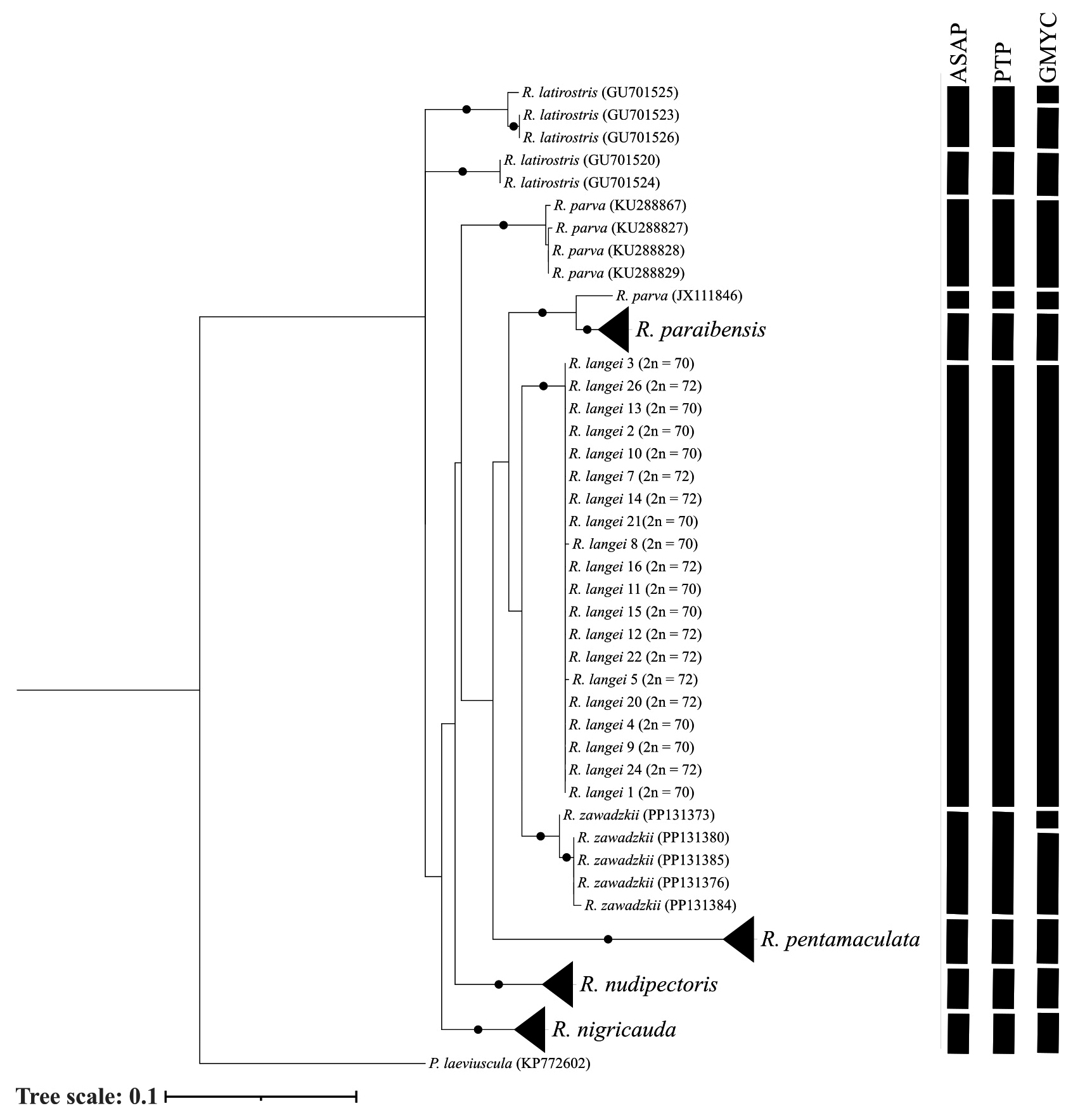
FIGURE 3| Gene tree constructed with sequences of the COI marker from Rineloricaria, using the maximum likelihood method with 1,000 bootstrap resamplings. The bars represent the results of the species delimiter analysis. Pseudoloricaria laeviuscula was used as an outgroup. Circles represent bootstrap values >80. Diploid numbers (2n = 70 and 2n = 72) are shown next to the corresponding terminal taxa of R. langei.
Discussion
This study identified numerical polymorphisms in R. langei, characterized by two distinct karyomorphs: one with 2n = 72 chromosomes (Karyomorph A) and another with 2n = 70 chromosomes (Karyomorph B). Despite differences in diploid number, both karyomorphs share an FN = 82, exhibit similar patterns of constitutive heterochromatin distribution, and display conserved localization of 18S and 5S rDNA sites. Remarkably, the observed diploid number of 72 chromosomes surpasses the previously established maximum for the genus (2n = 70). Given that 2n = 54 chromosomes is considered basal for the family Loricariidae (Artoni, Bertollo, 2001), centric fission likely represents a key evolutionary mechanism contributing to the increased diploid number observed in R. langei (2n = 70 and 72). According to Porto et al. (2014), structural chromosomal rearrangements, such as centric fissions, can elevate the diploid number without necessarily modifying the FN. This observation supports the pattern observed in R. langei, where an increase in 2n is accompanied by conservation of the FN.
Chromosomal polymorphism is a recurrent phenomenon in the genus Rineloricaria, characterized by intraspecific variation in both chromosomal number and structure. This pattern has been documented in R. lanceolata (Porto et al., 2014; Morais et al., 2024), R. latirostris (Glugoski et al., 2018, 2022), and R. lima (Rosa et al., 2012). In this study, we hypothesize that the observed chromosomal polymorphism, accompanied by conservation of the FN in R. langei, may have arisen through centric fissions occurring in a subtelocentric chromosome pair of karyomorph B (2n = 70; 2m + 10st + 58a), except the chromosome bearing the nucleolus organizer region. This process would result in an increase of four acrocentric chromosomes (from 58a to 62a) and a reduction of two subtelocentric chromosomes (from 10st to 8st), thereby generating the karyotype 2n = 72, composed of 2m + 8st + 62a, observed in karyomorph A.
Morais et al. (2024) suggest that chromosomal polymorphism in Rineloricaria may function as an adaptive mechanism that promotes genetic variability, particularly in small and isolated populations, a scenario frequently observed in species characterized by sedentary behavior and restricted geographic distributions. These chromosomal rearrangements can affect meiotic segregation and gene recombination, thereby fostering reproductive isolation and potentially facilitating speciation. Accordingly, the chromosomal rearrangements responsible for the observed karyotypic variation (2n = 70–72) in R. langei, which constitute a potential barrier to reproductive isolation and account for the absence of hybrids within a syntopic population, may be interpreted as evidence supporting cryptic speciation. While the molecular mechanisms underlying these processes remain incompletely elucidated, available evidence suggests that repetitive elements, including 5S and 18S rDNA, interstitial telomeric sequences, and hAT-type transposons (Glugoski et al., 2018), play a role in the origin and stabilization of mechanisms that elevate diploid number.
The terminal localization of NORs on the short arm of the first pair (pair 2) of subtelocentric chromosomes was demonstrated through silver nitrate staining and further substantiated by FISH utilizing an 18S rDNA probe in both karyomorphs. The presence of simple NORs, as described for R. langei, constitutes a conserved cytogenetic characteristic among major Rineloricaria species (Sassi et al., 2024). While the occurrence of multiple NORs has been reported in particular taxa, such as R. lanceolata from the Onça stream (Porto et al., 2014) and R. pentamaculata from the Tatupeba stream (Porto et al., 2011), these cases are considered derived exceptions. Such characteristics are generally attributed to the mobilization of transposable elements and ribosomal DNA transposition events.
The 5S rDNA signals in R. langei, which are confined to two acrocentric chromosome pairs (pairs 13 and 20), are congruent with the distribution patterns reported for several congeners. Nevertheless, the 5S rDNA marker exhibits considerable variability and evolutionary instability within the genus Rineloricaria. Cytogenetic analyses have revealed that certain species possess only two 5S rDNA sites, as observed in R. parva (Boulenger, 1895) (Takagui et al., 2020), and R. microlepidogaster (Regan, 1904)(Venturelli et al., 2021). Conversely, other species display pronounced amplification, with multiple 5S rDNA sites distributed across several chromosomes, as documented in R. capitonia Ghazzi, 2008, R. stellata Ghazzi, 2008, R. pentamaculata Langeani & de Araujo, 1994 (Primo et al., 2017; 2018; Cius et al., 2022), R. reisi Ghazzi, 2008 (Takagui et al., 2020), R. malabarbai Rodriguez & Reis, 2008, R. aequalicuspis Reis & Cardoso, 2001, R. quadrensis, R. longicauda (Venturelli et al., 2021), R. latirostris (Primo et al., 2017; Glugoski et al., 2018), and R. lima (Rosa et al., 2012). This heterogeneity is principally attributed to the terminal positioning of the 5S loci on subtelocentric or acrocentric chromosomal segments, which facilitates recurrent translocation events and recombination between non-homologous chromosomes (Primo et al., 2017). The pericentromeric localization of 5S rDNA in R. langei across both karyomorphs suggests a more conserved organizational pattern, characteristic of species exhibiting lower structural chromosomal dynamism. The observed stability of these loci in both karyomorphs further supports the notion that the differences in diploid chromosome number (70 and 72) did not compromise the integrity or chromosomal positioning of the minor ribosomal gene clusters, indicating that the chromosomal fissions involved in karyomorph differentiation did not directly affect chromosomes harboring 5S rDNA.
C-banding analysis in R. langei revealed a chromosomal pattern characteristic of the genus, consisting of prominent pericentromeric blocks of heterochromatin distributed across the majority of chromosomes, as well as a substantial heterochromatic block associated with the NOR on the first subtelocentric pair. This feature is notably pronounced in both observed karyomorphs (2n = 72 and 2n = 70). The co-localization of constitutive heterochromatin and the NOR has been extensively documented in Rineloricaria, as evidenced by studies on R. pentamaculata (Porto et al., 2014; Cius et al., 2022), R. lima (Rosa et al., 2012), R. malabarbai, R. cadeae (Hensel, 1868), R. microlepdogaster, R. aequalicuspis, R. quadrensis, and R. longicauda (Venturelli et al., 2021). These studies emphasize that the occurrence of a NOR accompanied by a substantial heterochromatic block constitutes a synapomorphic trait within the tribe Loricariini. The pronounced heterochromatic accumulation at the NOR in R. langei, particularly in the subtelocentric pair, aligns with this broader taxonomic pattern and may indicate a structural function in stabilizing the ribosomal chromosomal region and supporting the transcriptional activity of 18S rDNA genes.
In addition to cytogenetic data, molecular analyses provided further insights into the genetic variation within the studied population. The R. langei specimens analyzed presented three distinct haplotypes (H1, H2, and H3), with a maximum genetic divergence of 0.4%. According to the literature, COI gene divergence values below 1% are typically interpreted as intraspecific variation, whereas values exceeding 2% generally correspond to interspecific differentiation in Neotropical fishes (Hebert et al., 2003; Ward et al., 2005; Rossini et al., 2016). Thus, the genetic divergence values observed in this study fall below the established interspecific threshold, supporting the interpretation that the three haplotypes represent intraspecific variation within R. langei. Furthermore, the predominance of haplotype H1, detected in 18 of the analyzed individuals, indicates a high level of genetic homogeneity within the sampled population. This interpretation is further corroborated by the OTU delimitation analyses, in which ASAP, PTP, and GMYC consistently recovered R. langei as a single Operational Taxonomic Unit. The concordance among distance-based and tree-based approaches reinforces the absence of detectable species-level subdivision within the analyzed samples, despite the internal haplotypic variation.
Given the challenges associated with characterizing and identifying Rineloricaria species exclusively through morphological traits, the mitochondrial COI marker has emerged as a key molecular tool for delineating lineages and detecting cryptic diversity. Recent investigations, such as that by Castellanos-Mejía et al. (2024), have demonstrated the ability of COI to uncover independent evolutionary lineages, even within morphologically homogeneous groups, thereby reinforcing its value in integrative taxonomic frameworks and highlighting its pivotal role in the systematics of Rineloricaria. Recognizing the importance of broadening the molecular understanding of this genus, future research would be well served by incorporating additional molecular markers, which would facilitate a more comprehensive evaluation of genetic divergence among populations and species within Rineloricaria.
The phylogenetic reconstruction obtained in this study recovered R. langei and R. zawadzkii as sister lineages. This close relationship is compatible with their shared distribution in coastal drainages of the Serra do Mar and Paraíba do Sul (Silva et al., 2022) and may reflect recent divergence or incomplete lineage sorting rather than deep evolutionary separation; similar caveats concerning mitochondrial markers and the need for nuclear data have been previously noted (Mejía et al., 2023). Although the two species are morphologically distinct, notably in ventral plate coverage, morphological characters in Rineloricaria can be labile or homoplastic, so marked morphological differences do not necessarily contradict the observed mitochondrial affinity. Consequently, the sister-group pattern reinforces the need for broader geographic sampling and the inclusion of nuclear markers and cytogenetic data to resolve their evolutionary history (Silva et al., 2022; Mejía et al., 2023).
Within this context, the pattern observed in R. langei, marked by low mitochondrial divergence, a lack of consistent morphological differentiation, and distinct cytogenetic variation, aligns with scenarios in which molecular boundaries may only become apparent in later stages of speciation. When considered alongside the karyotypic stability in ribosomal regions and the subtle intrapopulational variation identified in the COI marker, these findings underscore the necessity of employing a multifaceted approach integrating cytogenetic, morphological, and multiple genetic markers to more accurately delineate evolutionary boundaries within the group. Overall, an integrative framework enhances the detection of markers capable of revealing subtle differentiation among recently diverged lineages, thereby elucidating patterns of evolutionary divergence that may remain undetected when using single-method approaches.
In summary, the findings indicate that cytogenetic and molecular data are both complementary and essential for the accurate taxonomic delimitation of R. langei. The presence of karyotypic variation alongside low mitochondrial divergence highlights the necessity of broadening geographic sampling and incorporating additional molecular markers to achieve a more comprehensive understanding of the evolutionary processes influencing this species. Consequently, the data produced in this study provide a robust foundation for future systematic, evolutionary, and conservation research in the Iguaçu River basin, while further emphasizing the importance of integrative approaches for elucidating the genetic and chromosomal dynamics within this genus.
References
Angulo A, Icochea J. Cryptic species complexes, widespread species and conservation: lessons from Amazonian frogs of the Leptodactylus marmoratus group (Anura: Leptodactylidae). Syst Biodivers. 2010; 8(3):357–70. https://doi.org/10.1080/14772000.2010.507264
Alves AL, Oliveira C, Foresti F. Karyotype variability in eight species of the subfamilies Loricariinae and Ancistrinae (Teleostei: Siluriformes: Loricariidae). Caryologia. 2003; 56(1):57–63. https://doi.org/10.1080/00087114.2003.10589308
Artoni RF, Bertollo LAC. Trends in the karyotype evolution of Loricariidae fish (Siluriformes). Hereditas. 2001; 134(3):201–10. https://doi.org/10.1111/j.1601-5223.2001.00201.x
Bertollo LA, Cioffi MB, Moreira-Filho O. Direct chromosome preparation from freshwater teleost fishes. In: Ozouf-Costaz C, editor. Fish cytogenetic techniques: ray-finned fishes and chondrichthyans. Boca Raton: CRC Press; 2015. p.21–26. https://doi.org/10.1201/b18534-4
Castellanos-Mejía MC, Londoño-Burbano A, Ochoa LE, García-Alzate CA, DoNascimiento C. Two new species of Rineloricaria (Siluriformes: Loricariidae) from trans-Andean rivers of Colombia, unveiled through iterative taxonomy. Ichthyol Herpetol. 2024; 112(3):429–43. https://doi.org/10.1643/i2023091
Cius A, Lorscheider CA, Barbosa LM, Zawadzki CH, Borin-Carvalho LA, Porto FE. Contributions of species Rineloricaria pentamaculata (Loricariidae: Loricariinae) in a karyoevolutionary context. Caryologia. 2022; 75(3):39–46. https://doi.org/10.36253/caryologia-1611
Drummond AJ, Suchard MA, Xie D, Rambaut A. Bayesian phylogenetics with BEAUti and the BEAST 1.7. Mol Biol Evol. 2012; 29(8):1969–73. https://doi.org/10.1093/molbev/mss075
Fricke R, Eschmeyer WN, Fong JD. Eschmeyer’s catalog of fishes: genera/species by family/subfamily. [Internet]. San Francisco: California Academy of Science; 2025a. Available from: http://researcharchive.calacademy.org/research/ichthyology/catalog/fishcatmain.asp
Fricke R, Eschmeyer WN, Van Der Laan R. Eschmeyer’s catalog of fishes: genera, species, references. [Internet]. San Francisco: California Academy of Science; 2025b. Available from: http://researcharchive.calacademy.org/research/ichthyology/catalog/fishcatmain.asp
Fichberg I. Relações filogenéticas das espécies do gênero Rineloricaria Bleeker, 1862 (Siluriformes: Loricariidae, Loricariinae). [PhD Dissertation]. São Paulo: Universidade de São Paulo; 2008. Available from: https://doi.org/10.11606/T.41.2008.tde-17022009-115207
Fujisawa T, Barraclough TG. Delimiting species using single-locus data and the generalized mixed Yule coalescent approach: a revised method and evaluation on simulated data sets. Syst Biol. 2013; 62(5):707–24. https://doi.org/10.1093/sysbio/syt033
Glugoski L, Giuliano-Caetano L, Moreira-Filho O, Vicari MR, Nogaroto V. Co-located hAT transposable element and 5S rDNA in an interstitial telomeric sequence suggest the formation of Robertsonian fusion in armored catfish. Gene. 2018; 650:49–54. https://doi.org/10.1016/j.gene.2018.01.099
Glugoski L, Nogaroto V, Deon GA, Azambuja M, Moreira-Filho O, Vicari MR. Enriched tandem repeats in chromosomal fusion points of Rineloricaria latirostris (Boulenger, 1900) (Siluriformes: Loricariidae). Genome. 2022; 65(9):479–89. https://doi.org/10.1139/gen-2022-0043
Hall TA. BioEdit: a user-friendly biological sequence alignment editor and analysis program for Windows 95/98/NT. Nucleic Acids Symp Ser. 1999; 41:95–98.
Hatanaka T, Galetti PM. Mapping of the 18S and 5S ribosomal RNA genes in the fish Prochilodus argenteus Agassiz, 1829 (Characiformes: Prochilodontidae). Genetica. 2004; 122:239–44. https://doi.org/10.1007/s10709-004-2039-y
Hebert PDN, Cywinska A, Ball SL, DeWaard JR. Biological identifications through DNA barcodes. Proc R Soc London Ser B Biol Sci. 2003; 270(1512):313–21. https://doi.org/10.1098/rspb.2002.2218
Howell WM, Black DA. Controlled silver-staining of nucleolus organizer regions with a protective colloidal developer: a 1-step method. Experientia. 1980; 36:1014–15. https://doi.org/10.1007/BF01953855
Ingenito LF, Ghazzi MS, Duboc LF, Abilhoa V. Two new species of Rineloricaria (Siluriformes: Loricariidae) from the rio Iguaçu basin, southern Brazil. Neotrop Ichthyol. 2008; 6(3):355–66. https://doi.org/10.1590/S1679-62252008000300009
Ivanova NV, Zemlak TS, Hanner RH, Hebert PDN. Universal primer cocktails for fish DNA barcoding. Mol Ecol Resour. 2007; 7(4):544–48. https://doi.org/10.1111/j.1471-8286.2007.01748.x
Kumar S, Stecher G, Tamura K. MEGA7: Molecular Evolutionary Genetics Analysis version 7.0 for bigger datasets. Mol Biol Evol. 2016; 33(7):1870–74. https://doi.org/10.1093/molbev/msw054
Levan A, Fredga K, Sandberg AA. Nomenclature for centromeric position on chromosomes. Hereditas. 1964; 52(2):201–20. https://doi.org/10.1111/j.1601-5223.1964.tb01953.x
Lui R, Blanco D, Moreira-Filho O, Margarido V. Propidium iodide for making heterochromatin more evident in the C-banding technique. Biotech Histochem. 2012; 87(7):433–38. https://doi.org/10.3109/10520295.2012.696700
Margarido VP, Moreira-Filho O. Karyotypic differentiation through chromosome fusion and number reduction in Imparfinis hollandi (Ostariophysi: Heptapteridae). Genet Mol Biol. 2008; 31(1):235–38. https://doi.org/10.1590/S1415-47572008000200012
Marajó L, Viana PF, Ferreira AMV, Rapp Py-Daniel LH, Cioffi MB, Sember A et al. Chromosomal rearrangements and the first indication of an ♀X1X1X2X2/♂X1X2Y sex chromosome system in Rineloricaria fishes (Teleostei: Siluriformes). J Fish Biol. 2023; 102(2):443–54. https://doi.org/10.1111/jfb.15275
Martins C, Galetti Jr. PM. Chromosomal localization of 5S rDNA genes in Leporinus fish (Anostomidae, Characiformes). Chromosome Res. 1999; 7:363–67. https://doi.org/10.1023/A:1009216030316
Mejía E, Ferraro GA, Buckup PA. A new species of Rineloricaria (Siluriformes: Loricariidae) from coastal drainages of Rio de Janeiro, southeastern Brazil. Neotrop Ichthyol. 2023; 21(1):e220083. https://doi.org/10.1590/1982-0224-2022-0083
Morais VIB, Oliveira JVL, Alesci A, Almeida MC, Artoni RF. Exploring chromosomal polymorphism and evolutionary implications in Rineloricaria lanceolata (Günther, 1868) (Siluriformes: Loricariidae): insights from meiotic behavior and phylogenetic analysis. Biology. 2024; 13(9):708. https://www.mdpi.com/2079-7737/13/9/708
Morais-Silva JP, Oliveira AV, Fabrin TMC, Diamante NA, Prioli SMAP, Frota A et al. Geomorphology influencing the diversification of fish in small-order rivers of neighboring basins. Zebrafish. 2018; 15(4):389–97. https://doi.org/10.1089/zeb.2017.155
Pereira LH, Hanner R, Foresti F, Oliveira C. Can DNA barcoding accurately discriminate megadiverse Neotropical freshwater fish fauna? BMC Genet. 2013; 14:20. https://doi.org/10.1186/1471-2156-14-20
Pinkel D, Straume T, Gray JW. Cytogenetic analysis using quantitative, high-sensitivity, fluorescence hybridization. PNAS. 1986; 83(9):2934–38. https://doi.org/10.1073/pnas.83.9.2934
Porto FE, Vieira MMR, Barbosa LM, Borin-Carvalho LA, Vicari MR, Portela-Castro ALB et al. Chromosomal polymorphism in Rineloricaria lanceolata Günther, 1868 (Loricariidae: Loricariinae) of the Paraguay basin (Mato Grosso do Sul, Brazil): evidence of fusions and their consequences in the population. Zebrafish. 2014; 11(4):318–24. https://doi.org/10.1089/zeb.2014.099
Primo CC, Glugoski L, Almeida MC, Zawadzki CH, Moreira-Filho O, Vicari MR et al. Mechanisms of chromosomal diversification in species of Rineloricaria (Actinopterygii: Siluriformes: Loricariidae). Zebrafish. 2017; 14:161–68. https://doi.org/10.1089/zeb.2016.138
Puillandre N, Brouillet S, Achaz G. ASAP: assemble species by automatic partitioning. Mol Ecol Resour. 2021; 21(2):609–20. https://doi.org/10.1111/1755-0998.13281
Rambaut A, Drummond AJ, Xie D, Baele G, Suchard MA. Posterior summarization in Bayesian phylogenetics using Tracer 1.7. Syst Biol. 2018; 67(5):901–04. https://doi.org/10.1093/sysbio/syy032
Rapp Py-Daniel LH. Phylogeny of the neotropical armored catfishes of the subfamily Loricariinae (Siluriformes: Loricariidae). [PhD Dissertation]. Tucson: The University of Arizona; 1997.
Rosa K, Ziemniczak K, Barros AV, Nogaroto V, Almeida MC, Cestari M et al. Numeric and structural chromosome polymorphism in Rineloricaria lima (Siluriformes: Loricariidae): fusion points carrying 5S rDNA or telomere sequence vestiges. Rev Fish Biol Fish. 2012; 22:739–49. https://doi.org/10.1007/s11160-011-9250-6
Rosenthal A, Coutelle O, Craxton M. Large-scale production of DNA sequencing templates by microtitre format PCR. Nucleic Acids Res. 1993; 21:173–74. https://doi.org/10.1093/nar/21.1.173
Rossini BC, Oliveira CAM, Melo FAG, Bertaco VA, Díaz de Astarloa JM, Rosso JJ et al. Highlighting Astyanax species diversity through DNA barcoding. PLoS ONE. 2016; 11:e0167203. https://doi.org/10.1371/journal.pone.0167203
Rosso JJ, Mabragaña E, González Castro M, Díaz de Astarloa JM. DNA barcoding Neotropical fishes: recent advances from the Pampa Plain, Argentina. Mol Ecol Resour. 2012; 12(6):999–1011. https://doi.org/10.1111/1755-0998.12010
Rozas J, Ferrer-Mata A, Sánchez-DelBarrio JC, Guirao-Rico S, Librado P, Ramos-Onsins SE et al. DnaSP 6: DNA sequence polymorphism analysis of large data sets. Mol Biol Evol. 2017; 34(12):3299–302. https://doi.org/10.1093/molbev/msx248
Sassi FMC, Cioffi MB, Moreira-Filho O. A state-of-art review of Loricariidae (Ostariophysi: Siluriformes) cytogenetics. Neotrop Ichthyol. 2024; 22(4):e240050. https://doi.org/10.1590/1982-0224-2024-0050
Silva GJC, Silva GSC, Oliveira C. A new species of spiny Rineloricaria (Siluriformes: Loricariidae) from the rio Paraíba do Sul basin and coastal rivers from Rio de Janeiro State. Zootaxa. 2022; 5175(2):285–92. https://doi.org/10.11646/zootaxa.5175.2.6
Soria TV, Fabrin TMC, Diamante NA, Mota TFM, Oliveira IJ, Oliveira AV et al. DNA barcoding analysis of Gymnotus species in two Neotropical river basins. Int Rev Hydrobiol. 2018; 103(5–6):120–26. https://doi.org/10.1002/iroh.201801954
Sumner AT. A simple technique for demonstrating centromeric heterochromatin. Exp Cell Res. 1972; 75(1):304–06. https://doi.org/10.1016/0014-4827(72)90558-7
Takagui FH, Baumgärtner L, Venturelli NB, Paiz LM, Viana P, Dionísio JF et al. Unrevealing the karyotypic evolution and cytotaxonomy of armored catfishes (Loricariinae) with emphasis in Sturisoma, Loricariichthys, Loricaria, Proloricaria, Pyxiloricaria, and Rineloricaria. Zebrafish. 2020; 17(5):319–32. https://doi.org/10.1089/zeb.2020.1893
Thompson JD, Higgins DG, Gibson TJ. Improving the sensitivity of progressive multiple sequence alignment through sequence weighting, position-specific gap penalties and weight matrix choice. Nucleic Acids Res. 1994; 22(22):4673–80. https://doi.org/10.1093/nar/22.22.4673
Venturelli NB, Takagui FH, Pompeo LRS. Cytogenetic markers to understand chromosome diversification and conflicting taxonomic issues in Rineloricaria (Loricariidae: Loricariinae) from Rio Grande do Sul coastal drainages. Biologia. 2021; 76:2561–72. https://doi.org/10.1007/s11756-021-00748-3
Ward RD, Zemlak TS, Innes BH, Last PR, Hebert PDN. DNA barcoding Australia’s fish species. Philos Trans R Soc Lond B Biol Sci. 2005; 360(1462):1847–57. https://doi.org/10.1098/rstb.2005.1716
Zhang J, Kapli P, Pavlidis P, Stamatakis A. A general species delimitation method with applications to phylogenetic placements. Bioinformatics. 2013; 29(22):2869–76. https://doi.org/10.1093/bioinformatics/btt499
Authors

Sérgio João Sartor Neto1, Ligia Carla Balini2, Lucas Pietro Ferrari Gianini3, Cláudio Henrique Zawadzki3,4, Fernanda Errero Porto5, Luciana Andreia Borin-Carvalho1,2, Alessandra Valéria de Oliveira1,4,6, Laura Ivana Ramos6 and Carlos Alexandre Fernandes1,3,4
[1] Departamento de Biotecnologia, Genética e Biologia Celular (DBC), Universidade Estadual de Maringá, Av. Colombo, 5790, 87020-900, Maringá, PR, Brazil. (SJSN) ra129260@uem.br, (LABC) labcarvalho@uem.br (AVO) avoliveira@uem.br, (CAF) cafernandes@uem.br (corresponding author).
[2] Programa de Pós-Graduação em Genética e Melhoramento, Universidade Estadual de Maringá, Av. Colombo, 5790, 87020-900, Maringá, PR, Brazil. (LCB) ligia_balini@hotmail.com.
[3] Programa de Pós-Graduação em Biologia Comparada, Universidade Estadual de Maringá, Av. Colombo, 5790, 87020-900, Maringá, PR, Brazil. (LPFG) luquindia@gmail.com, (CHZ) chzawadzki@nupelia.uem.br.
[4] NUPELIA – Núcleo de Pesquisas em Limnologia, Ictiologia e Aquicultura, Universidade Estadual de Maringá, Av. Colombo, 5790, 87020-900, Maringá, PR, Brazil.
[5] Departamento de Ciências Morfológicas, Universidade Estadual de Maringá, Av. Colombo, 5790, 87020-900, Maringá, PR, Brazil. (FEP) fepsaparolli@uem.br.
[6] Programa de Pós-Graduação em Ecologia de Ambientes Aquáticos Continentais, Centro de Ciências Biológicas (CCB), Universidade Estadual de Maringá, Maringá, PR, Brazil. (LIR) lauraivramos@gmail.com.
Authors’ Contribution 

Sérgio João Sartor Neto: Conceptualization, Data curation, Formal analysis, Investigation, Methodology, Writing-original draft, Writing-review and editing.
Ligia Carla Balini: Formal analysis, Investigation, Methodology, Writing-review and editing.
Lucas Pietro Ferrari Gianini: Formal analysis, Investigation, Methodology, Writing-review and editing.
Cláudio Henrique Zawadzki: Formal analysis, Investigation, Methodology.
Fernanda Errero Porto: Formal analysis, Investigation, Writing-review and editing.
Luciana Andreia Borin-Carvalho: Formal analysis, Investigation, Writing-review and editing.
Alessandra Valéria de Oliveira: Formal analysis, Investigation, Methodology, Writing-review and editing.
Laura Ivana Ramos: Formal analysis, Investigation, Methodology, Writing-review and editing.
Carlos Alexandre Fernandes: Project administration, Resources, Supervision, Validation, Visualization, Writing-review and editing.
Ethical Statement
The animals were captured with authorization from the Instituto Chico Mendes Institute de Conservação da Biodiversidade (ICMBio, number 94381–1). This study was carried out strictly following the recommendations of the Guide for the Care and Use of Laboratory Animals, approved by the Comitê de Ética em Experimentação Animal of the Universidade Estadual de Maringá (License number: 9877240524 – CEUA/UEM). The experiments followed ethical conduct, and before euthanasia, the fish were anesthetized with an overdose of clove oil (Griffiths, 2000).
Competing Interests
The author declares no competing interests.
Data availability statement
The authors confirm that the data supporting the findings of this study are available within the article.
AI statement
English language editing was performed using Grammarly (Grammarly Inc., USA).
Funding
Conselho Nacional de Desenvolvimento Científico e Tecnológico (CNPq), through the Pró-Reitoria de Pesquisa e Pós-Graduação of the Universidade Estadual de Maringá (UEM-PPG), for the scientific initiation scholarship granted to SJSN.
Supplementary Material
Supplementary material SUP
Peer Review
How to cite this article
Sartor Neto SJ, Balini LC, Gianini LPF, Zawadzki CH, Porto FE, Borin-Carvalho LA, Oliveira AV, Ramos LI, Fernandes CA. Integrative cytogenetic and molecular analyses reveal chromosomal polymorphism in Rineloricaria langei (Siluriformes: Loricariidae): reporting the highest diploid number in the genus. Neotrop Ichthyol. 2026; 24(2):e250214. https://doi.org/10.1590/1982-0224-2025-0214
Copyright
This is an open access article under the terms of the Creative Commons Attribution License, which permits use, distribution and reproduction in any medium, provided the original work is properly cited.
Distributed under
Creative Commons CC-BY 4.0

© 2025 The Authors.
Diversity and Distributions Published by SBI
 Accepted March 5, 2026
Submitted December 19, 2025
Epub July 20, 2026