![]() Laura M. Donin1
Laura M. Donin1 ![]() ,
, ![]() Juliano Ferrer2 and
Juliano Ferrer2 and ![]() Tiago P. Carvalho3
Tiago P. Carvalho3
PDF: EN XML: EN | Supplementary: S1 S2 S3 S4 S5 S6 S7 S8 S9 | Cite this article
Abstract
Cambeva contains species with complex taxonomy or poorly delimitated in terms of morphology and geopraphic distribution. We conducted an extensive review of Cambeva populations from coastal drainages of Southern to Southeastern Brazil to evaluate species geographic limits with an integrative analysis including morphological and molecular data (COI). We test if two single-locus methods, Bayesian Poisson Tree Processes (bPTP) and Generalized Mixed Yule Coalescent (GMYC), are efficient to delimit species boundaries in Cambeva by the comparison with the diagnosable morphological units. Using GMYC, we also evaluated the combination of tree and molecular clock priors to reconstruct the input phylogeny and assessed how well the implemented model fitted our empirical data. Eleven species were identified using a morphological diagnosability criterion: Cambeva balios, C. barbosae, C. botuvera, C. cubataonis, C. davisi, C. guaraquessaba, C. iheringi, C. tupinamba, and C. zonata and two treated as undescribed species. In contrast with previous knowledge, many of them have wider distribution and high intraspecific variation. Species delimitation based on single-locus demonstrated incongruences between the methods and strongly differed from the morphological delimitation. These disagreements and the violation of the GMYC model suggest that a single-locus data is insufficient to delimit Cambeva species and the failure may be attributable to events of mitochondrial introgression and incomplete lineage sorting.
Keywords: Color pattern,GMYC,Integrative taxonomy, Species delimitation, Trichomycterinae.
Cambeva contém espécies com taxonomia complexa ou mal delimitadas em termos morfológicos e de distribuição geográfica. Realizamos uma extensa revisão de populações de Cambeva das drenagens costeiras do Sul ao Sudeste do Brasil para avaliar os limites das espécies com uma análise integrativa incluindo dados morfológicos e moleculares (COI). Testamos se dois métodos de locus único, Implementação Bayesiana dos Processos da Árvore de Poisson (bPTP) e Coalescente de Yule Misto Generalizado (GMYC), são eficientes para delimitar os limites das espécies em Cambeva pela comparação com as unidades morfológicas diagnosticáveis. Usando o GMYC, também avaliamos a combinação de árvores e relógios moleculares para reconstruir a filogenia e avaliamos o quão bem o modelo implementado se ajustava aos nossos dados empíricos. Foram identificadas 11 espécies usando o critério morfológico: Cambeva balios, C. barbosae, C. botuvera, C. cubataonis, C. davisi, C. guaraquessaba, C. iheringi, C. tupinamba e C. zonata e duas tratadas como espécies não-descritas. Em contraste com o conhecimento prévio, muitas delas têm distribuição mais ampla e alta variação intraespecífica. A delimitação das espécies baseada em locus único demonstrou incongruências entre os métodos e diferiu fortemente da delimitação morfológica. Essas discordâncias e a violação do modelo GMYC sugerem que os dados de locus único são insuficientes para delimitar as espécies de Cambeva e a falha pode ser atribuída a eventos de introgressão mitocondrial e sorteio incompleto da linhagem.
Palavras-chave: Delimitação de espécie, GMYC, Padrão de coloração, Taxonomia integrativa, Trichomycterinae.
Introduction
The family Trichomycteridae contains 371 species (Fricke et al., 2022) distributed exclusively in the Neotropical region, the second richest Siluriformes family. The subfamily Trichomycterinae includes 270 species classified in nine genera, Bullockia Arratia, Chang, Menu-Marque & Rojas, 1978, Cambeva Katz, Barbosa, Mattos & Costa, 2018, Eremophilus Humboldt, 1805, Hatcheria Eigenmann, 1909, Rhizosomichthys Miles, 1943, Scleronema Eigenmann, 1917, Trichomycterus Valenciennes, 1832, Ituglanis Costa & Bockmann, 1993, and Silvinichthys Arratia, 1998; which represent more than two-thirds of the family diversity (Fricke et al., 2022). In terms of phylogenetic studies, the family has been investigated using anatomical systems [e.g., osteology (de Pinna, 1998) and musculature (Datovo, Bockmann, 2010)], and molecular data as the multi-locus approaches (Ochoa et al., 2017a; Henschel et al., 2018; Katz et al., 2018; Fernández et al., 2021) and with ultraconserved elements (Ochoa et al., 2020). As a result, there were significant advances in phylogeny and classification, such as establishing the monophyletic status of Trichomycterinae and rearrangements of its internal clades.
One of these clades units within Trichomycterinae was firstly namely “clade D4” by Ochoa et al. (2017a) and consists of Scleronema plus a subset of species of Trichomycterus, a long-standing “waste-basket” genus of Trichomycteridae (de Pinna, 1998). Posteriorly, Katz et al. (2018) proposed the genus Cambeva to allocate those species that formed a sister clade of Scleronema and currently contains 46 valid species (Fricke et al., 2022).
One of the puzzles to delimit trichomycterine species is associated with their variable intraspecific morphology. Color pattern, for example, is often used to diagnose Neotropical freshwater fishes but has been demonstrated to be labile for species of Cambeva and Trichomycterus due to aspects as ontogeny, geographic variation, or habitat specificity (see Arratia et al., 1978; Silva et al., 2010; Ferrer, Malabarba, 2013; Nascimento et al., 2017; Donin et al., 2020; Pereira et al., 2021). Besides that, the morphological variation of trichomycterine species is repeatedly poorly accessed, and some descriptions are conducted based solely on specimens from type localities (Costa, 1992; Triques, Vono, 2004; Wosiacki, Oyakawa, 2005; Barbosa, Costa, 2010; Costa et al., 2020a,b). Consequently, some of these species’ hypotheses need reevaluation after a careful examination of new samples, including ontogenetic series and broad geographic areas (Reis, de Pinna, 2019; DoNascimiento, Prada-Pedreros, 2020; Donin et al., 2020; Lima et al., 2021).
Single-locus sequence-based species delimitations have been increasingly used to resolve species limits in the diverse Neotropical fish fauna (Serrano et al., 2019; Agudelo-Zamora et al., 2020; Delapieve et al., 2020; Mateussi et al., 2020; Lima et al., 2021). In this regard, some methods using DNA sequences have an exploratory capacity, do not require previous information on species assignment, and are helpful in groups with uncertain and problematic alpha-taxonomy (Talavera et al., 2013; Luo et al., 2018). Currently, the most frequent tree-based methods applied to species delimitation are the Generalized Mixed Yule Coalescent (GMYC) and the Bayesian Poisson Tree Processes (bPTP), which either use branch lengths to identify branching rate transition points or the number of substitutions (Fujisawa, Barraclough, 2013; Zhang et al., 2013).
Although these methods have been recognized as a valuable tool to delimit species, critics regarding its methodological deficiencies, such as over splitting, limited information on single-locus analysis, and accuracy concerning different priors on inputted phylogenetic reconstructions (Talavera et al., 2013; da Cruz, Weksler, 2018). Additionally, their accuracy depends on a sampling of multiple individuals within each species, small effective population sizes, and the absence of population genetic structure within species (Fujisawa, Barraclough, 2013; Luo et al., 2018; Fonseca et al., 2021).
In this paper, an extensive review of Cambeva populations from coastal drainages of Southern to Southeastern Brazil was used to evaluate species limits in an integrative analysis with morphological and molecular data. The main question is whether single-locus methods, specifically GMYC and bPTP, are efficient to delimit species boundaries in the genus Cambeva. Additionally, we describe intraspecific variations and updated geographic distributions for the species of Cambeva in the study area and discuss their relevance facing previous taxonomic papers.
Material and methods
Study region and taxa sources. The study area ranges from Tramandaí River basin to small rivers in the São Paulo State, representing the known southern and northern limits, respectivaly of Cambeva in Brazilian coastal drainages (Fig. 1). We conducted a population level analysis (per basin) in the following drainages (south to north): Tramandaí River, Mampituba River, Araranguá River, Tubarão River, Cubatão Sul River, Biguaçu River, Tijucas River, Santa Catarina Island streams, Itapocu River, Cubatão Norte River, tributaries to Babitonga, Guaratuba and Paranaguá bays, Ribeira de Iguape River, and small coastal rivers of São Paulo State.
All fishes were anesthetized with a Eugenol solution to take pictures and extract tissue samples from muscle or fin clips. Photographs of live specimens were taken by TPC using the phototank immersion method proposed by Sabaj Pérez (2009). Posteriorly, the fishes were euthanized with a concentrated Eugenol solution (Lucena et al., 2013) and fixed in 10% formaldehyde. Currently, the specimens and the tissue samples are preserved in 70% and 96% ethanol, respectively, in the fish collection of the Departamento de Zoologia, Universidade Federal do Rio Grande do Sul, Porto Alegre (UFRGS). Additional specimens and tissue samples used in the study are listed in the Tab. S1 and material examined.

FIGURE 1| Geographical distribution of Cambeva species in the coastal drainages of Southern and Southeastern Brazil (crossed circles indicate their past known distribution). Some dots represent more than one locality.
Morphological data and morphospecies assignment. Primary species (or morphospecies) assignment was done based on a combination of morphological characters, typically used in the alpha-taxonomy of Cambeva (e.g., color pattern, morphometric, fin-ray counts, osteology, and pores of the laterosensory system), which enabled us to diagnose species within the study area. These characters were searched in the original descriptions (Ferrer, Malabarba, 2013;Costa et al., 2021; Bizerril, 1994; Haseman, 1911; Wosiacki, 2005; Eigenmann, 1917, 1918; Wosiacki, Oyakawa, 2005), newly taxonomic works (Wosiacki, 2005; Malabarba et al., 2013; Nascimento et al. 2017; Donin et al., 2020) or currently proposed. The following measurements were taken point to point with a digital caliper (precision 0.1 mm): (1) standard length, (2) head length, (3) head width, (4) pre-dorsal length, (5) pre-pelvic length, (6) pre-anal length, (7) scapular-girdle width, (8) trunk length, (9) pectoral-fin length, (10) pelvic-fin length, (11) distance between pelvic-fin base and anus, (12) caudal-peduncle length, (13) caudal-peduncle depth, (14) body depth, (15) dorsal-fin base length, (16) anal-fin base length, (17) snout length, (18) interorbital distance, and (19) eye diameter (Fig. 2); maxillary, nasal, and rictal barbels lengths (Tchernavin, 1944); mouth width and supraorbital pore s6 distance (Costa, 1992); and interopercular patch length (Ferrer et al., 2015). Principal component analysis (PCA) was applied to check the overall morphometric variation among populations and species with similar morphology and genetics, using measurements treated for size using a log-ratio transformation (Aitchinson, 1982). Osteological information was obtained from cleared and counterstained specimens (cs) according to procedures described by Taylor, Van Dyke (1985). Vertebrae counts exclude those of the Weberian complex, and the compound caudal centrum (PU1+ U1) was counted as a single element. Counts of unbranched and branched rays of the pectoral fin are represented by upper case Roman numeral and Arabic numerals, respectively. Nomenclature of the laterosensory canals and associated pores follows Rizzato, Bichuette (2016).

FIGURE 2| Schematic drawing of morphometric measurements applied to Cambeva species: (1) standard length, (2) head length, (3) head width, (4) predorsal length, (5) prepelvic length, (6) pre-anal length, (7) scapular girdle width, (8) trunk length, (9) pectoral-fin length, (10) pelvic-fin length, (11) distance between pelvic-fin base and anus, (12) caudal peduncle length, (13) caudal peduncle depth, (14) body depth, (15) length of dorsal-fin base, (16) length of anal-fin base, (17) snout length, (18) interorbital distance, and (19) eye diameter. Illustration made by Alexandre Ribeiro.
Molecular data and analyses. Genetic sequences used in the analyses are those cited by Donin et al. (2020; reference number: 7) in addition to others available in GenBank from coastal drainages and neighboring continental basins associated with the following papers: Silva et al. (2010; 1), Pereira et al. (2013; 2), Nascimento et al. (2017; 3), Ochoa et al. (2017a,b; 4), Katz et al. (2018; 5), and Morais-Silva et al. (2018; 6) (Tab. S1). In total, 170 sequences and 23 terminal taxa were used (Tab. S1). We adopted the taxonomic identification provided by these authors citing their authorship in the text and making their reference in the phylogenetic trees through the reference numbers mentioned above. The sequence identified as Trichomycterus paolence Eigenmann, 1917 (currently Cambeva; GenBank accession HM376398) from Pereira et al. (2013) do not align with any sequence of Cambeva and was not included in the analyses. The DNA extractions, PCR, sequences editing, and alignment followed Donin et al. (2020). We applied two different methods of species delimitation using fragments of the mitochondrial DNA cytochrome oxidase c subunit I (COI) to examine congruence between these and the delimitated morphospecies: Generalized Mixed Yule Coalescent (GMYC; Fujisawa, Barraclough, 2013; Pons et al., 2006) and Bayesian Poisson tree processes (bPTP; Zhang et al., 2013). The methods differ in that GMYC uses branch lengths (timed divergences) to identify when divergence times more closely resemble coaslescence events on a time-calibrated ultrametric tree, while in PTP the method model speciation by using the number of substitutions, and a ultrametric tree is not required (Zhang et al., 2013).
The COI alignment was partitioned by codon position to determine the best models and partition schemes of molecular evolution in PartitionFinder v.1.1.1 (Lanfear et al., 2012) under the Bayesian information criterion (BIC). To conduct the GMYC analysis, we assess the impact of four different combinations of priors [clock models (strict and relaxed lognormal) and tree priors (yule and a coalescent prior of constant population size)] on the delimitation of species (defined here as combination 1, 2, 3 and 4). Using these combinations of distinct priors (clock model and tree prior) we estimated an ultrametric tree, because the model requires an ultrametric tree as input (Fujisawa, Barraclough, 2013), in BEAST v.2.5.0 (Drummond et al., 2012), programmed to run for 40 million generations (MCMC), sampling every 4 x 10³ generations. The convergence was assessed by assuring that all parameters reached stationarity and effective sample sizes (i.e., ESS > 200) using Tracer v.1.7.1 (Rambaut et al., 2018). The first 4 million generations (10%) were discarded as burn-in of 10%, and the remaining trees were used to summarize the results of the Bayesian analysis, using the Maximum Clade Credibility Tree (MCC) in TreeAnnotator BEAST v.1.7 (Rambaut, Drummond, 2013). The MCC was checked in FigTree v1.4.3 and used as an input file (Newick format) for the GMYC analyses performed in the GMYC server (https://species.h-its.org/gmyc/) under a single threshold method. We also assessed the statistical fit of the GMYC model using the parametric bootstrap (100 replications) approach of the R package P2C2M.GMYC (Fonseca et al., 2021) for all the four inputted phylogenetic trees that used a distinct combination of priors in BEAST. P2C2M.GMYC calculates the number of species clusters for the empirical dataset then uses the number of inferred species to simulate a Yule tree and coalescent genealogies for each terminal lineage using the number of individuals inferred to be in each species cluster. Then simulate sequences and a UPGMA tree for each tree simulated under the correct model and compare the empirical and simulated number of species after replicates. This procedure is suggested to be included in the analytical pipeline of GMYC analyses (Fonseca et al., 2021).
In the Bayesian Poisson tree processes (bPTP), we generated a non-ultrametric best maximum likelihood (ML) tree implemented in IQ-TREE web server (http://iqtree.cibiv.univie.ac.at/) (Trifinopoulos et al., 2016) under an Ultrafast Bootstrap analysis (Hoang et al., 2018) and the remaining of the parameters with default values. The best model for nucleotide substitution was estimated under a Bayesian criterion (BIC) in IQ-TREE web server (http://iqtree.cibiv.univie.ac.at/). The bPTP analysis was performed in the bPTP server (https://species.h-its.org/ptp/) under default values.
Collection abbreviations. CPUFMT, Coleção de Peixes da Universidade Federal do Mato Grosso, Cuiabá; DZSJRP, Coleção de Peixes da Universidade Estadual Paulista Júlio de Mesquita Filho, São José do Rio Preto; FMNH, Division of Fishes, Department of Zoology, Field Museum of Natural History, Chicago; MCN, Museu de Ciências Naturais (ex-Fundação Zoobotânica do Rio Grande do Sul), SEMA-RS, Porto Alegre; MCP, Museu de Ciências e Tecnologia da Pontifícia Universidade Católica do Rio Grande do Sul, Porto Alegre; MNRJ, Museu Nacional, Rio de Janeiro; MPEG, Museu Paraense Emílio Goeldi, Zoologia, Laboratório de Ictiologia, Belém; MZUEL, Museu de Zoologia da Universidade Estadual de Londrina, Londrina; MZUSP, Museu de Zoologia da Universidade de São Paulo, São Paulo; NUP, Coleção Ictiológica do Nupélia, Universidade Estadual de Maringá, Maringá; UNICTIO, Laboratório de Ictiologia, Universidade do Vale do Rio dos Sinos, São Leopoldo.
Results
Morphospecies identity and geographic distribution. We recognized 11 morphospecies of Cambeva across the study area (Fig. 1; Tab. 1). Nine of them can be associated with valid species names: Cambeva balios (Ferrer & Malabarba, 2013), C. barbosae Costa, Feltrin & Katz, 2021, C. botuvera Costa, Feltrin & Katz, 2021, C. cubataonis (Bizerril, 1994), C. davisi (Haseman, 1911), C. guaraquessaba (Wosiacki, 2005), C. iheringi (Eigenmann, 1917), C. tupinamba (Wosiacki & Oyakawa, 2005), and C. zonata (Eigenmann, 1918), and two are treated as undescribed species (Cambeva sp. 1 and Cambeva sp. 2).
In the study area, C. balios (Fig. 3A) occurs exclusively in the headwaters of the Mampituba River basin. Cambeva balios is distinguished from its congeners in the coastal basins, except C. botuvera, by the coloration with two distinct layers in adult specimens composed of large rounded black blotches in the inner layer and small spots in the outer layer over a pale-yellow background (vs. body with mottled pattern of blotches not rounded, with spots or rounded blotches restricted to form a mid-lateral line, or with small circular blotches not coalescent). It differs from C. botuvera by having 10–13 ventral procurrent caudal-fin rays (vs. 14–15).
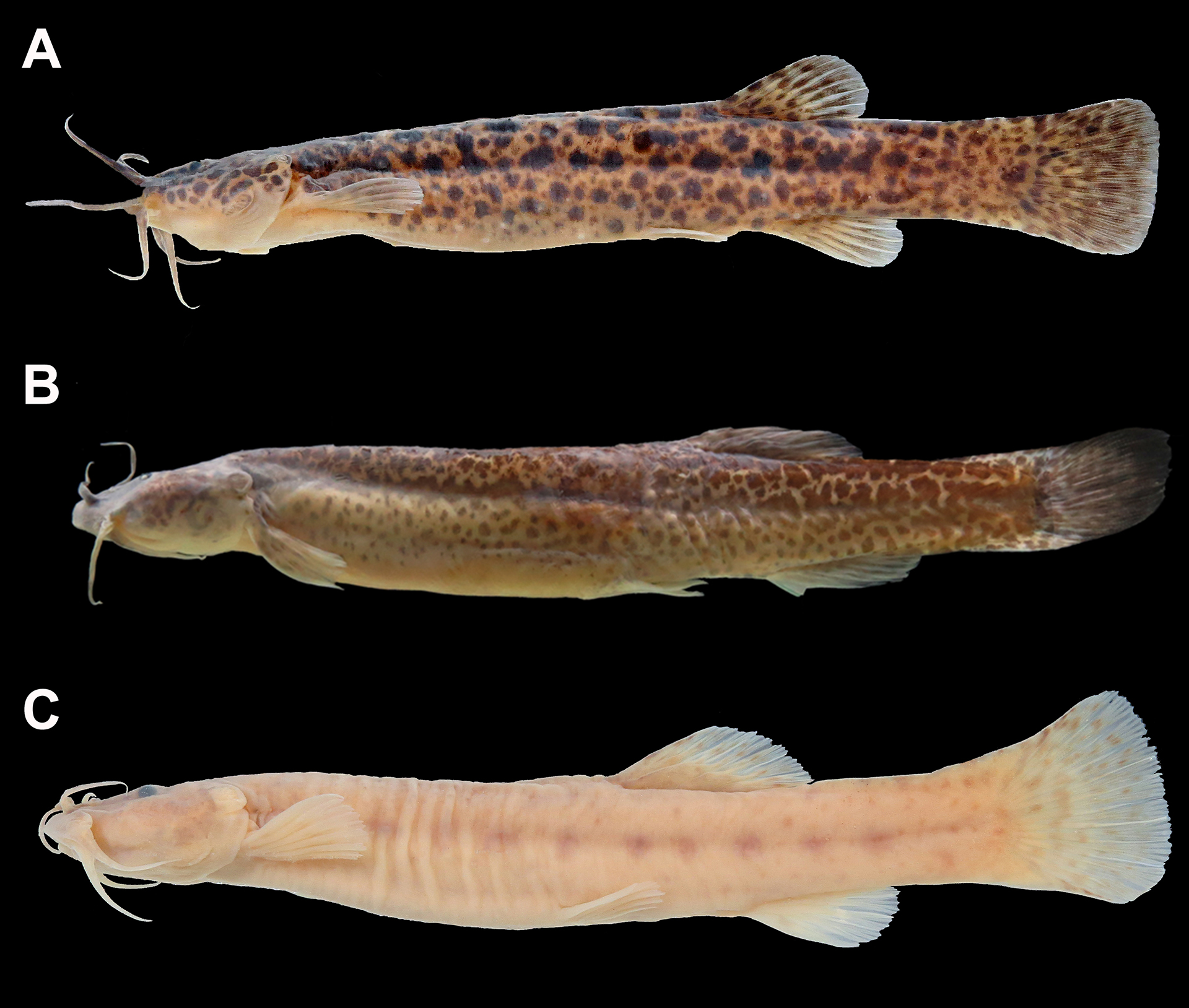
FIGURE 3| A. Cambeva balios from Mampituba River basin, UFRGS 16295, 63.8 mm SL. B. Cambeva davisi from Ribeira de Iguape River basin, MZUEL 17202, 72.8 mm SL, fixed in alcohol. C. Cambeva iheringi from Piagui River, coastal drainage of São Paulo State, MNRJ 24008, 75.6 mm SL.
TABLE 1 | Primary species (or morphospecies) recognized along the study area and their diagnostic morphological characters typically used in the alpha-taxonomy of Cambeva from coastal basins of South and Southeast Brazil.
Morphospecies/characters | Pectoral-fin
rays: modal number | Dorsal
procurrent caudal-fin rays: number | Ventral
procurrent caudal-fin rays: number | Branchiostegal
rays: number | Vertebrae:
number | Ribs:
number | First
pterygiophore of dorsal fin position: anterior to neural spine of vertebra | First
pterygiophore of anal fin position: anterior to hemal
spine of vertebra | Antorbital
segment of the infraorbital canal (pores ‘i1’, ‘i3’) | Presumably
adults body: overall coloration |
Cambeva balios | I,6 | 14–16 | 12 | 9 | 39–40 | 12–13 | 20–21 | 22–23 | absent | Two distinct layers composed of large
rounded black blotches in the inner layer and small spots in the outer layer
over a pale yellow background |
Cambeva barbosae | I,7 | 17–22 | 10–12 | 7–8 | 36–37 | 11–12 | 16–18 | 21–22 | absent | Mottled pattern composed of irregular
and coalescent inconspicuous black or with sparse black blotches varying in,
eventually rounded, over a pale yellow background |
Cambeva botuvera | I,6 | 17–19 | 14–15 | 8 | 39–40 | 12–14 | 20–21 | 23 | absent | Two distinct layers composed of large
ellipsoid black blotches, sometimes coalescent and forming a mid-lateral
stripe, vermicular black marks, or yet inconspicuous black spotted in the
inner layer; and small spots in the outer layer over a pale
yellow background |
Cambeva cubataonis | I,6 | 17–21 | 12–13 | 9 | 38–40 | 12–15 | 18–20 | 22–24 | usually present | Mottled pattern composed of coalescent
black blotches sometimes forming saddle marks dorsally, over a pale yellow background or almost completely black with
thin yellowish areas |
Cambeva davisi | I,6 | 17 | 11 | 9–10 | 37–38 | 13–15 | 19 | 22–23 | absent | Mottled pattern composed of coalescent
and irregular black marks over a pale yellow
background |
Cambeva guaraquessaba | I,7 | 15 | 8 | 8 | 37 | 14–15 | 18 | 22 | present | Light brown background with a
mid-lateral line composed of black spots with about the eye size |
Cambeva iheringi | I,7 | 18 | 8 | 7–8 | 35 | 11 | 15–16 | 21–22 | present | Numerous black spots or sparse rounded
black blotches, sometimes forming a mid-lateral line, over a grayish or a pale yellow background |
Cambeva tupinamba | I,7 | 15–16 | 10 | 7–8 | 39–40 | 14–15 | 19–20 | 23–25 | present | Brown background with a mid-lateral
black stripe along the trunk and sparse black spots |
Cambeva zonata | I,6 | 13–15 | 9–12 | 8–9 | 37 | 13 | 18–19 | 21 | absent | Mottled pattern composed of coalescent
black marks, sometimes forming a mid-lateral stripe over a pale
yellow background, and never with saddle marks dorsally |
Cambeva sp. 1 | I,5 | 14–17 | 10–12 | 8–9 | 36–39 | 12–14 | 17–20 | 20–23 | absent | Mottled pattern composed of coalescent
and irregular black marks over a pale yellow
background |
Cambeva sp. 2 | I,6 | 17–21 | 12–14 | 7–8 | 38–40 | 14–15 | 19 | 22–23 | absent | Pale yellow background with small
circular black blotches not coalescent |
Cambeva barbosae (Fig. 4) is distributed in the Cubatão Sul, Biguaçu, Tijucas, Itapocu, Cubatão Norte river basins and in the Santa Catarina Island. Cambeva barbosae is distinguished from its congeners by the combination of a high number of pectoral-fin rays modally I,7 (vs. I,5 or I,6), the antorbital segment of the laterosensory system (pores i1 and i3) absent (vs. present), and by the coloration with a mottled pattern composed of irregular and coalescent inconspicuous black marks (Figs. 4A, B) or with sparse black blotches varying in size (Figs. 4C, D), eventually rounded (Fig. 4E) (vs. body with large rounded or ellipsoid black blotches; with spots or rounded blotches forming a mid-lateral line, with small circular blotches not coalescent).

FIGURE 4| Polymorphic color pattern within of Cambeva barbosae ranging from a mottled color pattern in individuals from (A) Santa Catarina Island, UFRGS 23183, 80.1 mm SL, and (B) Biguaçu River basin, UFRGS 22907, 61.3 mm SL; to a blotched color pattern with dark marks varying in shape and size from Biguaçu River basin, (C, D) UFRGS 22932, 58.7mm SL and 51.0 mm SL, respectively, and (E) UFRGS 20937, 30.0 mm SL.
Cambeva botuvera (Fig. 5) is widely distributed in the Itajaí River basin being distinguished from other congeners in the coastal area, except C. balios, by the coloration with two distinct layers in adult specimens composed of large ellipsoid black blotches (Figs. 5A, F), sometimes coalescent and forming a mid-lateral stripe (Figs. 5B, D), vermicular black marks (Fig. 5E), or yet inconspicuous black-spotted (Fig. 5C) in the inner layer; and small spots in the outer layer over a pale yellow background (vs. body with mottled pattern of blotches not ellipsoid, with spots or rounded blotches restricted to form a mid-lateral line, or with small circular blotches not coalescent). It differs from C. balios by having 14–15 ventral procurrent caudal-fin rays (vs. 10–13).

FIGURE 5| Variation on the coloration in vivo and ontogenetic series of Cambeva botuvera from (A, C–F) Itajaí-Açu River, UFRGS 23182, 47.3 mm SL, 64.8 mm SL, 68.1 mm SL, 81.1 mm SL, 95.6 mm SL, respectively; and (B) from Itajaí-Mirim River, UFRGS 24558, 67.1 mm SL, showing the color pattern with two layers in the skin composed of large and small round black blotches in the inner and outer layer, respectively.
Cambeva cubataonis (Fig. 6) is distributed in the Itapocú River, Cubatão Norte River, and tributaries of Babitonga, Guaratuba and Paranaguá bays. Cambeva cubataonis is distinguished by the antorbital segment of the laterosensory system (pores i1 and i3) usually present (variable only in a few individuals from Babitonga and Guaratuba bays) (vs. always absent), the number of pectoral-fin rays modally I,6 (vs. I,5 or I,7), and coloration with a mottled pattern composed of coalescent black blotches sometimes forming saddles dorsally, over a pale-yellow background (Figs. 6A, B) or almost entirely black with thin yellowish areas (vs. body with mottled pattern not forming saddles dorsally; with large rounded or ellipsoid black blotches; with spots or rounded blotches forming a mid-lateral line, or with small circular blotches not coalescent)(Figs. 6C, D).

FIGURE 6| Variation on the coloration in Cambeva cubataonis, showing a mottled color pattern composed by blotches variable in shape and forming marked saddles dorsally, (C–E) specimens with large dark areas merged giving an overall dark color pattern. Specimens from tributaries of Guaratuba Bay (A) UFRGS 24549, 53.1 mm SL; (B) UFRGS 24556, 60.3 mm SL; and from tributaries to Babitonga Bay (C–E) UFRGS 24552, 77.2 mm SL, 85.7 mm SL, and 61.6 mm SL, respectively.
Cambeva davisi (Fig. 3B) is restricted to the headwaters of the Ribeira de Iguape River basin being distinguished from its congeners by its mottled color pattern composed of coalescent and irregular black marks (vs. mottled pattern forming saddles dorsally; body with large rounded or ellipsoid black blotches; with spots or rounded blotches forming a mid-lateral line, with small circular blotches not coalescent) the first anal-fin pterygiophore placed anteriorly to the 22nd‒23rd hemal spines of the vertebral column (vs. anteriorly to the 20‒25th), and the antorbital segment of the laterosensory system (pores i1 and i3) absent (vs. present).
Cambeva iheringi (Fig. 3C) is known from coastal drainages along the Serra do Mar escarpment in the São Paulo State. Cambeva iheringi is distinguished from its congeners by the low number of vertebrae (35 vs. 36‒40) and its coloration composed of numerous black spots or sparse rounded black blotches, sometimes concentrated along de mid-lateral line of the body, over a pale-yellow or a grayish background (vs. body with mottled pattern of blotches).
Cambeva guaraquessaba (Figs. 7A, B) occurs exclusively in the tributaries to the Paranaguá Bay, being distinguished from its congeners by the color pattern with a mid-lateral line composed of black spots with about the eye size (Figs. 8A, B) (vs. body with blotches or spots not forming a mid-lateral stripe), 37 vertebrae (vs. 35‒40), and the first dorsal- and anal-fin pterygiophores placed anteriorly to the 18th neural spine and 22nd hemal spine of the vertebral column, respectively (vs. anteriorly to the 16th‒21st and 20‒25th, respectively).
Cambeva tupinamba (Figs. 7C, D) occurs exclusively in the Ribeira de Iguape River basin being distinguished from its congeners by the color pattern with a series of spots forming anteriorly (to the anal-fin origin) a mid-lateral black stripe along the trunk (Figs. 7C, D) (vs. body with blotches or spots not forming a mid-lateral stripe), 39 to 40 vertebrae (vs. 35‒40), the first dorsal- and anal-fin pterygiophores placed anteriorly to the 19th‒20th neural spine and 23th‒25th hemal spine of the vertebral column, respectively (vs. anteriorly to the 15th‒21st; 20‒24th, respectively).
Cambeva zonata (Fig. 8) is restricted to the Ribeira de Iguape River basin along the Betari River and Iporanga River sub-basins. Cambeva zonata is distinguished by its mottled color pattern composed of coalescent black marks, sometimes forming a mid-lateral stripe in juveniles, but with no clear saddled pattern (vs. body with mottled pattern forming saddles dorsally; with large rounded or ellipsoid black blotches; with spots or rounded blotches forming a mid-lateral line, or with small circular blotches not coalescent); the first anal-fin pterygiophore placed anteriorly to the 21th hemal spine of the vertebral column (vs. anteriorly to the 22nd‒24th), and the antorbital segment of the laterosensory system (pores i1 and i3) absent (vs. present).

FIGURE 7| Coloration pattern of Cambeva species. (A, B). Cambeva guaraquessaba, UFRGS 24554, 37.0 and 36.9 mm SL, respectively, tributary of Guaraqueçaba River basin, Paranaguá Bay. C–D. Cambeva tupinamba, UFRGS 24550, 63.0 mm SL and 39.4 mm SL, Betari River, Ribeira de Iguape River basin.
In the coastal region, Cambeva sp. 1 (Fig. 9) is the southernmost morphospecies inhabiting the Tramandaí River system (specifically the Maquiné and Três Forquilhas river basin), Mampituba, and Araranguá river basins. Cambeva sp. 1 is distinguished from its congeners by the lower number of pectoral-fin rays (modally I,5 vs. I,6 or I,7).
Cambeva sp. 2 (Fig. 10) is distributed in the Araranguá and Tubarão river basins and differs from its congeners by a color pattern composed of circular black blotches, about eye size or slightly larger, that are not coalescent (body with mottled pattern of coalescent blotches, with spots or rounded blotches restricted to form a mid-lateral line). Additionally, Cambeva sp. 2 is distinguished from C. balios and C. botuvera (both eventually have a similar color pattern in juveniles) by the higher number of dorsal procurrent caudal-fin rays (17‒21 vs. 10‒16), and the first dorsal-fin pterygiophore placed anteriorly to the 19th neural spine of the vertebral column (vs. anteriorly to the 20th‒21st).

FIGURE 8| Coloration pattern of Cambeva zonata, (A–C) UFRGS 24538, (A) 51.1 mm SL, (B) 48.4 mm SL, Betari River, Ribeira de Iguape River basin, and (C) 41.4 mm SL.
Given the morphological and genetic similarity between the populations of Cambeva balios from the Laguna dos Patos System (type locality) and the Mampituba River basin with C. botuvera (see below) from the Itajaí River basin, we promote a morphometric comparison. A PCA showed that these populations overlap to some degree (Fig. S2). More precisely, the populations of C. balios from Laguna dos Patos and C. botuvera from the Itajaí River basin overlap at a higher level than the population of C. balios from the Mampituba River basin. This fact is mainly explained by the high values of PC1 presented in the C. balios population from Mampituba River basin associated with longer barbels length (i.e., nasal, maxillary, and rictal). In sum, PC1 explains 44.69% and PC2 20.23% of the variation observed in the analyzed specimens.

FIGURE 9| Individuals representing populations of Cambeva sp. 1 from distinct river drainages. (A) Maquiné River basin, UFRGS 22211, 58.6 mm SL, (B) Mampituba River basin, MCP 23623, 55.7 mm SL; and (C, D) Araranguá River basin, UFRGS 22962, 45.2 and 59.8 mm SL, respectively.
Cambeva guaraquessaba and C. tupinamba are genetically similar (see below), but the PCA (Fig. S2) showed a separation between them associated mainly with the supra-orbital pore distance. PC1 explains 32.70% and PC2 22.45% of the variation observed in the analyzed specimens.
Phylogenetic reconstruction. We investigated the phylogenetic relationships of Cambeva species using a mitochondrial matrix of COI (597 bp) under a Bayesian inference. The best models for nucleotide substitution estimated by codon position in PartitionFinder for the BEAST analyses were TrNef + I, HKY and GTR+G, respectively. The phylogenetic analysis of Cambeva (Fig. 11; Fig. S7) resulted in three large species-inclusive clades with relative high support (posterior probability values – PP): named here in as clade “A” (PP = 0.99), clade “B” (PP = 0.94), and clade “C” (PP = 1). The clade A includes a subclade (PP = 0.99) containing C. brachykechenos (Ferrer & Malabarba, 2013), C. poikilos (Ferrer & Malabarba, 2013), C. perkos (Datovo, Carvalho & Ferrer, 2012),and Cambeva sp. 1 sister-group to another subclade (PP = 0.41) composed by C. balios, C. botuvera, C. diatropoporos (Ferrer & Malabarba, 2013), and C. tropeiro (Ferrer & Malabarba, 2011). In clade A, samples belonging to the morphologically delimited species C. balios, C. botuvera, C. perkos, and Cambeva sp. 1 were recovered as non-monophyletic. Cambeva balios plus C. botuvera form a well-supported monophyletic group sister-group to C. tropeiro (PP = 1).

FIGURE 10| Variation on the coloration of Cambeva sp. 2 from (A–D) Tubarão River, UFRGS 24553, 66.1, 45.4, and 56.04, 52.1 mm SL, and (E, F) Araranguá River, UFRGS 22964, 61.1 and 60.7 mm SL, respectively, showing a unique color pattern that differs from other congeners in the study area: one layer of coloration composed of not coalescent round small blotches.
The Clade B includes C. barbosae, C. cubataonis, C. davisi, C. diabola (Bockmann, Casatti & de Pinna, 2004), C. flavopicta Costa, Feltrin & Katz, 2020, C. horacioi Reis, Frota, Fabrin & Graça, 2019, C. naipi (Wosiacki & Garavello, 2004), C. pascuali (Ochoa, Silva, Costa e Silva, Oliveira & Datovo, 2017), C. perkos, C. stawiarski (Miranda Ribeiro, 1968), C. zonata, and Cambeva sp. 2. Internal relationships in Clade B are generally poorly resolved (low supports) and several species are not monophyletic, including those from coastal drainages, excepting Cambeva sp. 2, which has two populations (Araranguá and Tubarão river basins) reciprocally monophyletic (PP = 1). Populations of C. barbosae are nested within C. cubataonis sensu Katz et al. (2018) and C. diabola sensu Pereira et al. (2013) (PP = 0.6), being sister group to C. davisi sensu Nascimento et al. (2017) plus C. perkos sensu Donin et al., 2020 (PP = 1). Specimens representing C. cubataonis form a monophyletic group (PP = 1) excluding the likely misidentified specimens of C. cubataonis sensu Katz et al. (2018). Cambeva zonata is nested within a lineage along with Cambeva davisi sensu Morais-Silva et al. (2018). Cambeva davisi were recovered as polyphyletic, appearing independently in several portions of the clade B. Specifically, the population of C. davisi (sensu Nascimento et al., 2017) from the Ribeira de Iguape River basin is inserted in a lineage with Cambeva stawiarski sensu Donin et al. (2020) and C. davisi sensu Ochoa et al. (2017a) and Morais-Silva et al. (2018) from upper Paraná and Iguaçu river basins (PP = 1).
The Clade C contains C. iheringi, C. guaraquessaba, C. tupinamba, C. variegata (Costa, 1992), which were recovered as non-monophyletic except for the latter with a single sample in the analysis. Cambeva guaraquessaba and C. tupinamba form a highly supported monophyletic group (PP = 1) sister-group of a clade of C. iheringi represented by samples from continental and coastal drainages (PP = 0.96).
Species delimitation and congruence. The mitochondrial COI (597 bp), was used for the species delimitation analyses (GMYC, bPTP; S1). Best-fit models of nucleotide substitution estimated for the non-ultrametric for bPTP tree were TIM+F+I+G4. We conducted the GMYC analysis using distinct ultrametric reconstruction in BEAST resulted from a combination of distinct priors (combinations 1, 2, 3, and 4; i.e., clock models and tree priors).
The GMYC results differed significantly depending on the prior combination used for the phylogenetic reconstruction (Fig. 11; Tab. 2; Figs. S3‒S7), and none was substantially congruent with the morphological assessment. In these analyses, 2, 5, 5, and 43 groups were suggested (Tab. 2; Figs. S3‒S7) with large confidence intervals in the number of clusters.
Combination 1 (relaxed clock lognormal/yule model) has only two clusters (ranging from 1 to 23; Tab. 2; Fig. S3), groups all species of clade A in one cluster and all species from clades B and C on another. Combination 2 (relaxed clock -lognormal /coalescent constant population size) supports five cluster (from 2 to 45; Tab. 2; Fig. S4). This combination groups clade A into 2 clusters: the first formed by Cambeva tropeiro and C. balios, and the second by C. brackykechenos, C. diatropoporos, C. perkos (Datovo, Carvalho & Ferrer, 2012), C. poikilos, and the undescribed species Cambeva sp. 1; clade B into two clusters, one containing Cambeva sp. (= C. sp.; two juvenile specimens with uncertain morphological identification), C. davisi, C. flavopicta, C. horacioi, C. pascuali, C. taroba (Wosiacki & Garavello, 2004), C. zonata, and Cambeva sp. 2; another with C. barbosae, C. cubataonis, C. davisi, C. diabola (Bockmann, Casatti & de Pinna, 2004), C. perkos and C. stawiarski; and the last cluster formed by species from clade C (C. guaraquessaba, C. iheringi, and C. tupinamba). Cambeva naipi is suggested as a single entity.
Combination 3 (strict clock/yule model) presents the same groupings of the combination 2 (ranging from 1 to 43; Tab. 2; Fig. S5), except for Cambeva sp. 2, which groups with C. barbosae, C. cubataonis, C. davisi, C. diabola, C. naipi, C. perkos,and C. stawiarski.

FIGURE 11| Bayesian phylogenetic tree of Cambeva speciesobtained from COI data. The vertical red line shows the coalescent branching process estimated by using the single-threshold model in the GMYC combination 4 (strict clock model vs. a coalescent constant population size tree prior).
Combination 4 (strict clock/coalescent constant population size) is the one that generated the largest number of clusters (43 with confidence interval from 5 to 46; Tab. 2; Figs. S6 and S7). Likelihoods for the cutoff point in the GMYC analyses were similar throughout most of the diversification of Cambeva in all combinations examined (Tab. 2). Fitting of the empirical data on the GMYC model using the parametric bootstrap test of the P2C2M. GMYC analysis indicates that the model was violated (p-value = 0; threshold = 0.1) in three of the four phylogenetic reconstructions using the distinct priors. The GMYC model was not violated (p-value = 0.99; threshold = 6.7) only in the analysis where the input tree was reconstructed using a combination of a strict molecular clock and a coalescent tree prior of coalescent constant population size (Fig. 11; Figs. S6 and S7).
TABLE 2 | Comparative results of GMYC species delimitation for each Clock/Prior combination tested.
Combinations Clock
Model/Prior | 1 | 2 | 3 | 4 |
Relaxed/Yule | Relaxed/CCP | Strict/Yule | Strict/CCP | |
Likelihood of null model | 804.0655 | 835.278 | 800.5291 | 834.1315 |
Maximun likelihood of GMYC model | 804.5015 | 838.5136 | 802.0595 | 839.2084 |
Likelihood ratio | 0.8719561 | 6.471159 | 3.060767 | 10.15375 |
Number of ML clusters | 2 | 5 | 5 | 43 |
Confidence interval | 1-23 | 2-45 | 1-43 | 5-46 |
Number of ML entities | 2 | 6 | 5 | 67 |
Confidence interval | 1-169 | 2-94 | 1-169 | 5-84 |
Threshold time | -1.470165 | -1.664397 | -1.364488 | -0.08717104 |
The PTP results support the presence of 13 clusters and 8 single entities to the maximum likelihood (mPTP) (Fig. S8); and average 35 clusters and 77 single entities, respectively, for the Bayesian analysis (bPTP) (Fig. S9). The estimated number of species ranges from 61 to 129, with a mean of 98. Most of the PTP results only partially corroborate the morphological delimitation of the coastal species of Cambeva. The mPTP corroborates only the morphologically delimited species Cambeva sp. 1 and Cambeva sp. 2 (Fig. S8). The bPTP results support either clustering of morphologically delimitated species (e.g., C. zonata with C. davisi) or splitting within the same morphospecies (e.g., Cambeva sp.1) (Fig. S9). These two analyses are largely incongruent (Figs. S8 and S9).
Discussion
Species delimitation methods. Popular methods of species delimitation based on single-locus data like bPTP and GMYC are widely used in biodiversity assessments in the Neotropical ichthyofauna (García-Melo et al., 2019; Agudelo-Zamora et al., 2020;Delapieve et al., 2020, Mateussi et al., 2020). We recognize that these methods can be a crucial exploratory tool in recognizing species. However, it must be done with caution. Our results indicated that, based on the dataset used, the species delimitations within Cambeva were highly variable between the used methods and mostly incongruent with the morphological delimitation. Within the GMYC, we also tested the impact of the different priors on grouping morphospecies (Talavera et al., 2013; Luo et al., 2018), resulting in different boundaries on each prior tested, ranging widely from 2 to 43 clusters. These results go against what was found by Talavera et al. (2013) that the results did not indicate substantial differences in the number of clusters found using input trees recovered by Coalescent or Yule priors and relaxed or strict molecular clocks.
The evaluation of the statistical fit of GMYC method shows that the model was violated in most of the phylogenetic reconstructions using the distinct priors (except for the combination of strict clock model with a coalescent constant population size tree prior). Simulation testing demonstrates that the underlying process of diversification and effective population size influences how well the GMYC model fits a given dataset (Luo et al., 2018; Fonseca et al., 2021). At small values of effective population size, allele coalescence occurs faster than speciation, being straightforward to model the differences between these processes. Taking geographic distribution on distinct and isolated basins as a proxy for effective population size, Cambeva shows a combination of widespread species presenting genetically structured populations in isolated basins (e.g., C. barbosae, C. cubataonis, Cambeva sp. 1, Cambeva sp. 2) but also species restricted to small areas in single basins (e.g., C. tropeiro and C. zonata). Other demographic scenarios that affect GMYC model fit are population expansion and geographic structure within species. In Cambeva’ species, faunal interchange between coastal and continental basins may trigger rapid population expansion (e.g., Cambeva sp. 1).
In addition to the violation of the GMYC model, the disagreement between the methods tested suggests that the analysis used here based on single-locus data is likely not adequate to assess the species limits within Cambeva. This failure can also be attributable to the intrinsic COI polymorphism properties, such as overlap in inter- and intraspecific divergences and non-monophyly of the species likely because of introgression or incomplete lineage sorting) (Knowles, Carstens, 2007; Talavera et al., 2013). Additionally, the performance of these single-locus-based methods strongly decreases when population sizes and speciation rates are high (Dellicour, Flot, 2015), and, in these cases, multilocus approaches are recommended. For the GMYC model, for example, a certain lumping tendency has been demonstrated and attributed to the poorly model ability to work process as incomplete lineage sorting or clades undergoing rapid radiation (Esselstyn et al., 2012; Reid, Carstens, 2012; Talavera et al., 2013). In our dataset, such a tendency was observed in the clade C with lumping between C. guaraquessaba and C. tupinamba in both GMYC and bPTP analyses.
Currently, studied introgression (hybridization) cases between species or genetically structured populations are rare in Neotropical fishes (e.g., Willis et al., 2012 in Cichla Bloch & Schneider, 1801; Sales et al., 2018 in Prochilodus Agassiz, 1829; Barreto et al., 2020 in Nematocharax Weitzman, Menezes & Britski, 1986). Events of hybridization were proposed between genetically structured populations of Nematocharax among isolated river basins in coastal rivers of Eastern Brazil (Barreto et al., 2020). These events were associated with capture stream events of populations in the headwaters of these basins. This type of hybridization event associated with rearrangement of drainage basins may be the case of Cambeva populations that inhabit headwaters and often occur in sympatry.
Inferences regarding species boundaries based on genetic data alone need to be analyzed carefully, and we reinforce those inferences of species boundaries should be conducted with consideration of other data sources, such as morphology, in an integrative approach (Pereira et al., 2013; Camelier et al., 2018; Serrano et al., 2019). Additionally, we suggest considering factors that can affect the efficiency of the delimitation method used for a given dataset, and we reinforce the need to test the dataset and models used in these analyzes (i.e., Fonseca et al., 2021), which is rarely done in these species delimitation analyses.
In this sense, we observe in the literature that incongruence across the results from different species delimitation methods are relatively common, and inferences and taxonomical acts drawn from these results are often not conservative. Thereby, in most contexts, it is better to fail to delimit species than it is to falsely delimit entities that do not represent independent evolutionary lineages (Carstens et al., 2013).
Cambeva taxonomy and distribution. Trichomycterinae is the most species-rich group of Trichomycteridae with 270 species, and by far, the one with more recent additions, with 93 species described in the last decade (Fricke et al., 2022). Several new species have been proposed in Trichomycterinae in the last years, but some species remain poorly defined. One of the caveats is that species are described based on a few specimens (e.g., < 10) and from limited localities, many of them, known from the type locality only (Wosiacki, 2005; Costa et al., 2020a,b; Katz, Costa, 2020), limiting assessment of intraspecific morphological and molecular variation. Contemplating a wide geographic sampling range and specimens of varied sizes are the main recommendations to properly investigate intraspecific color variation (Menezes et al., 2015; Nascimento et al., 2017), which deserves to be highlighted. Specifically, in Trichomycterinae, this feature has been extensively demonstrated to be associated with ontogeny (Castellanos-Morales, 2007; Lima et al., 2008; Ferrer, Malabarba, 2013; Nascimento et al., 2017); microhabitat preference (Arratia et al., 1978; Arratia, Menu-Marque, 1981; Arratia, 1983) or both (Silva et al., 2010).
The species richness of Cambeva has also increased substantially, with more than half (26) of the 46 valid species described in the past ten years. Cambeva is a dominant genus among the restricted-range species from Brazil (Nogueira et al., 2010) with many species restricted to the upper portions of drainages. In fact, our review demonstrated that there are restricted-range examples along the study area (e.g., C. guaraquessaba and C. zonata), but the most species have wider distribution than previously supposed (Fig. 1). However, the most notable distribution extensions were demonstrated in species recently described or redescribed: Cambeva barbosae to Tijucas and Cubatão Norte basins; C. botuvera to a wide area along the Itajaí River basin; and C. cubataonis to Babitonga, Guaratuba, and Paranaguá bays. Curiously, these species have the highest levels of color variation in the study area (Figs. 4‒6; Tab. 1), which also were poorly explored in their descriptions. In addition, C. barbosae and C. cubataonis are sympatric in the Itapocu and Cubatão Norte river basins, a condition not noticed by Costa et al. (2021) and Katz, Barbosa (2014).
The coloration of C. barbosae and C. cubataonis are usually similar, with the former composed of irregular and coalescent inconspicuous black marks or sparse blotches (Fig. 4) and the latter ranging from a mottled pattern of coalescent black blotches to a body almost entirely black (Fig. 6). For now, we assume that these color variations are not associated with ontogeny since we evaluate individuals of varied sizes, but preferences and association of individuals or populations with specific microhabitats deserve further investigation (Donin et al., 2020).
The intraspecific variation in the coloration is even more pronounced in C. botuvera, which has two distinct color layers in adult specimens composed of large ellipsoid black blotches, sometimes coalescent and forming a mid-lateral stripe, vermicular black marks, or yet inconspicuous black-spotted in the inner layer, and small spots in the outer layer (Fig. 5). Cambeva balios also has two distinct layers of coloration with similar variations (see Ferrer, Malabarba, 2013) and the only diagnostic characters from C. botuvera are the number of procurrent rays (Tab. 1). Considering these often-intraspecific variable morphological differences between the two species in addition to their almost indistinguishable coloration with layers of rounded blotches and genetic similarity, the validity of C. botuvera needs a more extensive evaluation.
The current morphological delimitation provided was conducted based on the diversity of Cambeva found in the study area and the species diagnosis are restricted to a sample of characters (Tab. 1). Given that, the present work does not aim to propose new diagnosis or recommend their broad usage within the genus. In the meantime, we recognize that some diagnosis, descriptions, and geographic delimitation for the taxa analyzed are current insufficient and a taxonomic review in the area in progress. In addition, considering all incongruences in the species delimitation methods and the high intraspecific variability found in Cambeva’ species, we reinforce the search and combination of multiples evidences to support hypotheses of species in this complex group.
Material examined. Lots and information in addition to those cited in Donin et al. (2020) and Ferrer, Malabarba (2020).“c&s” = specimens cleared and counterstained; “tis” = samples tissues followed by their collection codes. All from Brazil. Cambeva balios:Taquari-Antas River basin. UFRGS 16233, 3 (1 c&s), paratypes, 47.1–81.9 mm SL; UFRGS 16341, 3 (1 c&s), paratypes, 35.2–67.2 mm SL. Uruguai River basin: UFRGS 21886, 9 (tis TEC 6861 A, B), 19.6–74.4 mm SL. Mampituba River basin. UFRGS 16295, 10 (2 c&s), 48.6–80.1 mm SL; UFRGS 16299, 2, 41.3–60.8 mm SL; UFRGS 16284 (tis TEC 2738 A, B), 2, 27.5–28.5 mm SL; UFRGS 17198 (tis TEC 3203), 1, 59.4 mm SL. Cambeva barbosae:Cubatão Sul River basin. MCP 16570, 1, 32.4 mm SL; MCP 17477, 2, 31.9–57.5 mm SL; UFRGS 22906, 17 (tis TEC 7344 A, B; 2 c&s), 33.5–70.6 mm SL; UFRGS 24296, 6, 28.1–56.2 mm SL. Biguaçu River basin. UFRGS 22907, 2 (tis TEC 7345 A, B), 36.5–61.3 mm SL; UFRGS 22932, 6 (tis TEC 7360 A, B; 1 c&s), 36.5–66.5 mm SL. Tijucas River basin. UFRGS 20914, 1, 50.9 mm SL;UFRGS 20915,2 (1c&s),41.1–49.8 mm SL; UFRGS 23235, 1 (tis TEC 7458), 24.3 mm SL. Santa Catarina Island. UFRGS 23183, 4 (tis TEC 7447 A, B), 49.1–80.0 mm SL. Itapocu River basin. MCP 32214, 3, 57.5–62.9 mm SL; MCP 32190, 1, 44.9 mm SL; MNRJ 38421, 1, 63.1 mm SL; MNRJ 52565, 2, 45.3–47.3 mm SL; NUP 14240, 1, 53.5 mm SL; UFRGS 18481, 1, 54.1 mm SL; UFRGS 28701, 3, 43.5–51.2 mm SL. Cubatão Norte River basin: MCP 50476, 1, 52.0 mm SL; MCP 50485, 1, 42.6 mm SL; MNRJ 13191, 1, 39.1 mm SL; MNRJ 40952, 7, 43.4–52.6 mm SL; UFRGS 28698, 4, 41.3–71.2 mm SL; UFRGS 28697, 2 (1 c&s), 44.1–53.6- mm SL; UFRGS 28699, 27 (1 c&s), 22.7–54.1 mm SL. Cambeva botuvera: Itajaí River basin. UFRGS 20118, 8, 25.2–111.9 mm SL; UFRGS 22939 (tis TEC 7365 A, E; 2 c&s), 25, 37.9–78.8 mm SL; UFRGS 23182 (tis TEC 7446 A, C, F, I, L, M, P; 1 c&s), 27, 33.6–104.4 mm SL; UFRGS 24527, 1, 68.4 mm SL; UFRGS 24558, 4, 47.4–65.5 mm SL. Cambeva brackykechenos: Sinos River basin. UFRGS 24666, 1 (tis TEC 8229), 43.3 mm SL. Cambeva cubataonis:Itapocu River basin. MNRJ 41029, 1, 66.4 mm SL; UFRGS 24449, 1, 68.4 mm SL. Cubatão Norte River basin. UFRGS 24311, 2 (tis TEC 7996 A), 47.18–86.05 mm SL; UFRGS 24543, 6 (tis TEC 8117 A; 1 c&s), 32.7–76.8 mm SL; UFRGS 24567, 2 (1 c&s), 51.5–55.5 mm SL. Babitonga Bay. UFRGS 24552, 4 (tis TEC 8126 A, B; 1 c&s), 61.6–85.7 mm SL. Guaratuba Bay. UFRGS 24549, 8 (1 c&s), 53.1–64.7 mm SL; UFRGS 24556, 5 (tis TEC 8130 A, B), 45.3–62.9 mm SL. Paranaguá Bay:MNRJ 32940, 1 (tis MNLM 4644), 57.0 mm SL. Cambeva davisi:Upper Paraná River basin. MZUEL 11776, 10 (2 c&s), 30.8–92.2 mm SL. Iguaçu River basin. MZUEL 14788 (tis A, B), 2, 42.4-55.9 mm SL. Ribeira de Iguape River basin. MZUEL 17202 (tis A, B), 4, 38.3–73.1 mm SL; MZUEL 17213 (tis A, B), 2, 45.4–50.5 mm SL. Cambeva diatropoporos: Taquari-Antas River basin. UFRGS 17887, 1 (tis TEC 3560), 59.1 mm SL; UFRGS 17889, 1 (tis TEC 3562), 34.2 mm SL. Cambeva flavopicta: Uruguai River basin. MCP 17440, 5 (1 c&s), 29.9–61.5 mm SL; UFRGS 3964, 1, 29.9 mm SL; UFRGS 14998 (tis TEC 1780 A, B), 2, 28.7–30.2 mm SL; UFRGS 15000, 15, 21.6–42.2 mm SL. Cambeva guaraquessaba:Paranaguá Bay. MNRJ 40846, 11 (tis 7152), 53.8–36.5 mm SL; UFRGS 24541, 1 (tis TEC 8115 A), 30.88 mm SL; UFRGS 24554, 4 (tis TEC 8128 A), 39.4–46.7 mm SL. Cambeva iheringi: MNRJ 20129, 3, 36.3–44.2 mm SL. Cambeva naipi: Iguaçu River basin. UFRGS 11405, 1 (tis TEC 6739 A), 42.2 mm SL. Cambeva pascuali: Paranapanema River basin. MZUSP 121681, holotype, 49.1 mm SL. Cambeva perkos:Paranapanema River basin. MZUSP 82372, 1 paratype, 74.4 mm SL.Ijuí River basin UFRGS 17923 (tis TEC 3596 A), 1, 58.9 mm SL; UFRGS 18252 (tis TEC 3749 A), 1, not measured. Cambeva poikilos: Jacuí River basin. UFRGS 15014, 1 (tis TEC 1795 A), 56.3 mm SL; UFRGS 20352, 1 (tis TEC 5622 A), 60.2 mm SL. Sinos River basin. UFRGS 17419, 1 (tis TEC 3314 A), 59.2 mm SL; Taquari-Antas River basin. UFRGS 24604, 1 (tis TEC 8177 A), 54.3 mm SL. Cambeva stawiarski: Iguaçu River basin. UFRGS 18307, 10 (tis TEC 3793 A, B; 2 c&s), 31.7–49.4 mm SL. Cambeva taroba: Iguaçu River basin. UFRGS 18308, 1 (tis TEC 3794 A), not measured. Cambeva tupinamba:Ribeira de Iguape River basin. MZUSP 83727, 2, 61.0–64.4 mm SL; UFRGS 25094, 2, 52.8–70.4 mm SL. UFRGS 24550, 2 (tis TEC 8124 A, B), 39.8–62.1 mm SL; UFRGS 24563, 2 (tis TEC 8137 A, B), 22.7–43.4 mm SL. Cambeva tropeiro: Antas River. UFRGS 21878, 2 (tis TEC 6855 A, B), 81.1–83.2 mm SL. Cambeva zonata: Ribeira de Iguape River basin. UFRGS 18675, 1 (tis TEC 3998 A), 48.2 mm SL; UFRGS 24538, 11 (tis TEC 8112 A, B), 47.1–61.6 mm SL. Cambeva sp. 1:Tramandaí River basin. MCN 18587, 1, 62.7 mm SL; MCP 25375, 1, 47.0 mm SL; MCP 25395, 1, 52.0 mm SL; MCP 29117, 1, 46.1 mm SL; MCP 29143, 1, 32.2 mm SL; MCP 29147, 1, 38.6 mm SL; MCP 29694, 1, 20.7 mm SL; UFRGS 10651, 1 (c&s), 66.5 mm SL; UFRGS 17575, 1, 58.60 mm SL; UFRGS 19149, 2 (tis TEC 4867 A, B), not measured; UFRGS 19169, 1 (c&s), 61.4 mm SL; UFRGS 19178, 1 (tis TEC 4881 A), 55.3 mm SL; UFRGS 21346, 1, 52.3 mm SL; UFRGS 22211, 1, 58.6 mm SL; UFRGS 23494, 1 (tis TEC 7583 A), 53.0 mm SL. Mampituba River basin. MCP 23623, 2; 51.7–55.7 mm SL; UFRGS 17220, 2 (tis TEC 3225 A, B), 51.0–55.2 mm SL; UFRGS 17228, 1 (tis TEC 3233 A), 46.6 mm SL; UNICTIO 906, 1, 60.2 mm SL. Araranguá River basin. MCP 10646, 5, 51.6–20.4 mm SL; MZUEL 07547, 1, 39.2 mm SL; UFRGS 22962, 6 (2 tis TEC 7378 A, B; 1 c&s), 27.7–59.8 mm SL; UNICTIO 2460, 2, 53.2-60.8 mm SL. Cambeva sp. 2: Tubarão River basin. CPUFMT 79, 11, 12.7–18.5 mm SL; CPUFMT 86, 2, 22.6–28.3 mm SL; CPUFMT 106, 1, 36.1 mm SL; UFRGS 23184, 2 (tis TEC 7448 A, B), 31.2–51.8 mm SL; UFRGS 23185, 3 (tis TEC 7449 A; 1c&s), 33.8–59.8 mm SL; UFRGS 24553, 12 (1 c&s), 25.9-65.7 mm SL. Araranguá River basin. MCP 10642, 10, 28.0–49.6 mm SL; UFRGS 22964, 7 (tis TEC 7380 A, B; 2 c&s), 41.2–61.0 mm SL. Itajaí River basin: UFRGS 22939, 2 (tis TEC 7365 F, I), 35.3– 45.1 mm SL. Ituglanis amphipotamus:Ribeira de Iguape River basin. MZUSP 69393, holotype, 72.5 mm SL. Ituglanis australis:Uruguay River basin. MZUSP 112505, holotype, 56.4 mm SL. Ituglanis bambui:Tocantins River basin. MZUSP 79860, holotype, 42.1 mm SL. Ituglanis boitata: Tramandaí River basin. UFRGS 18455, holotype, 102.9 mm SL. Tubarão River basin. UFRGS 22903, 3, 45.8–63.4 mm SL; UFRGS 24547, 3, 58.3–91.7 mm SL. Ituglanis goya:Tocantins River basin. MZUSP 119759, holotype, 66.8 mm SL. Ituglanis inusitatus: Uruguai River basin. UFRGS 21829, holotype, 62.2 mm SL. Trichomycterus astromycterus:Doce River basin. MZUSP 124559, holotype, 51.1 mm SL. MZUSP 123760, 5 paratypes, 33.2-52.3 mm SL. Trichomycterus dali: Paraguai River basin. MZUSP 106630, holotype, 76.9 mm SL. Trichomycterus itacarambiensis:São Francisco River basin. MZUSP 50548, 4 paratypes, 21.1–47.3 mm SL. Trichomycterus jacupiranga:Ribeira de Iguape River basin. UFRGS 24555, 2 (tis TEC 8129 A, B), 35.1–44.8 mm SL. Trichomycterus payaya:Itapicuru River basin. MZUSP 88164, 4, paratypes, 33.5–36.3 mm SL.
Acknowledgments
We thank the personal and respective institutions for sending specimens and help during museum visits. We are grateful to labmates from UFRGS for the help in several steps of this work and Juliana Wingert (UFRGS) in scientific collection procedures. For support provided in field expeditions, Nathália Pio and Priscila Madoka Ito (UFRGS). Emília Wendt for help and suggestions on the molecular procedures. Fernando R. Carvalho and Kátia Tognini for all support. Alexandre Ribeiro for the Figs. 2 and 11, and Mateus Camana for the Fig. 1. This study was financed by the Coordenação de Aperfeiçoamento de Pessoal de Nível Superior – Brasil (CAPES) – Finance Code 001 (LMD – Master Degree and TPC –PNPD postdoctoral fellowship); JF was supported by CNPq with a postdoctoral scholarship and is currently supported by a PNPD fellowship (process #88887.463771/2019-00). The field expeditions of this project were funded by a CNPq Universal grant (#401204/2016-2) to L. R. Malabarba, of which are collaborators.
References
Agudelo-Zamora HD, Tavera J, Murillo YD, Ortega-Lara A. The unknown diversity of the genus Characidium (Characiformes: Crenuchidae) in the Chocó biogeographic region, Colombian Andes: Two new species supported by morphological and molecular data. J Fish Biol. 2020; 97(6):1662–75. https://doi.org/10.1111/jfb.14527
Aitchinson J. The statistical analysis of compositional data. J R Stat Soc B. 1982; 44(2):139–60. https://doi.org/10.1111/j.2517-6161.1982.tb01195.x
Arratia G. Trichomycterus chungaraensis n. sp. and Trichomycterus laucaensis n. sp. (Pisces, Siluriformes, Trichomycteridae) from the high Andean range. Stud Neotrop fauna E. 1983; 18(2):65–87.
Arratia G, Menu-Marque S. Revision of the freshwater catfishes of the genus Hatcheria (Siluriformes, Trichomycteridae) with commentaries on ecology and biogeography. Zool Anz. 1981; 207(1–2):88–111.
Arratia G, Chang A, Menu-Marque S, Rojas G. About Bullockia gen. nov., Trichomycterus mendozensis n. sp. and revision of the family Trichomycteridae (Pisces, Siluriformes). Stud Neotrop fauna E. 1978; 13(3–4):157–94.
Barbosa MA, Costa WJEM. Seven new species of the catfish genus Trichomycterus (Teleostei: Siluriformes: Trichomycteridae) from southeastern Brazil and redescription of T. brasiliensis. Ichthyol Explor Freshw. 2010; 21(2):97–122.
Barreto SB, Knowles LL, Affonso PRAM, Batalha-Filho H. Riverscape properties contribute to the origin and structure of a hybrid zone in a Neotropical freshwater fish. J Evolution Biol. 2020; 33(11):1530–42. https://doi.org/10.1111/jeb.13689
Bizerril CRSF. Descrição de uma nova espécie de Trichomycterus (Siluroidei, Trichomycteridae) do Estado de Santa Catarina, com uma sinopse da composição da família Trichomycteridae no leste Brasileiro. Arq Biol Tecnol. 1994; 37(3):617–28.
Camelier P, Menezes NA, Costa-Silva GJ, Oliveira C. Molecular and morphological data of the freshwater fish Glandulocauda melanopleura (Characiformes: Characidae) provide evidences of river captures and local differentiation in the Brazilian Atlantic Forest. PLoS ONE. 2018; 13(3):e0194247. https://doi.org/10.1371/journal.pone.0194247
Carstens BC, Pelletier TA, Reid NM, Satler JD. How to fail at species delimitation. Mol Ecol. 2013; 22(17):4369–83. https://doi.org/10.1111/mec.12413
Castellanos-Morales CA. Trichomycterus santanderensis: A new species of troglomorphic catfish (Siluriformes, Trichomycteridae) from Colombia. Zootaxa. 2007; 1541(1):49–55. https://doi.org/10.11646/zootaxa.1541.1.5
Costa WJEM. Description de huit nouvelles espèces du genre Trichomycterus (Siluriformes: Trichomycteridae), du Brésil oriental. Revue Française d’Aquariologie et Herpétologie. 1992; 18(4):101–10.
Costa WJEM, Feltrin CRM, Katz AM. A new species from subtropical Brazil and evidence of multiple pelvic fin losses in catfishes of the genus Cambeva (Siluriformes, Trichomycteridae). Zoosyst Evol. 2020a; 96(2):715–22. https://doi.org/10.3897/zse.96.56247
Costa WJEM, Mattos JLO, Amorim PF, Vilardo PJ, Katz AM. Relationships of a new species support multiple origin of melanism in Trichomycterus from the AtlanticForest of south-eastern Brazil (Siluriformes: Trichomycteridae). Zool Anz. 2020b; 288:74–83. https://doi.org/10.1016/j.jcz.2020.07.004
Costa WJEM, Feltrin CRM, Katz AM. Filling distribution gaps: Two new species of the catfish genus Cambeva from southern Brazilian Atlantic Forest (Siluriformes, Trichomycteridae). Zoosyst Evol. 2021; 97(1):147–59. https://doi.org/10.3897/zse.97.61006
da Cruz MOR, Weksler M. Impact of tree priors in species delimitation and phylogenetics of the genus Oligoryzomys (Rodentia: Cricetidae). Mol Phylogenet Evol. 2018; 119:1–12. https://doi.org/10.1016/j.ympev.2017.10.021
Datovo A, Bockmann FA. Dorsolateral head muscles of the catfish families Nematogenyidae and Trichomycteridae (Siluriformes: Loricarioidei): comparative anatomy and phylogenetic analysis. Neotrop Ichthyol. 2010; 8(2):193–246. http://dx.doi.org/10.1590/S1679-62252010000200001
Delapieve ML, Carvalho TP, Reis RE. Species delimitation in a range-restricted group of cascudinhos (Loricariidae: Epactionotus) supports morphological and genetic differentiation across coastalrivers of southern Brazil. J Fish Biol. 2020; 97(6):1748–69. https://doi.org/10.1111/jfb.14538
Dellicour S, Flot J-F. Delimiting species-poor data sets using single molecular markers: a study of barcode gaps, haplowebs and GMYC. Syst Biol. 2015; 64(6):900–08. https://doi.org/10.1093/sysbio/syu130
DoNascimiento C, Prada-Pedreros S. A new troglomorphic species of Trichomycterus (Siluriformes: Trichomycteridae) from north-eastern Colombia, with proposal of a new Trichomycterus subclade and remarks on some nominal species from Colombia. J Fish Biol. 2020; 96(4):968–85. https://doi.org/10.1111/jfb.14286
Donin LM, Ferrer J, Carvalho TP. Taxonomical study of Trichomycterus (Siluriformes: Trichomycteridae) from the Ribeira de Iguape River basin reveals a new species recorded in the early 20th century. J Fish Biol. 2020; 96(4):886–904. https://doi.org/10.1111/jfb.14278
Drummond AJ, Suchard MA, Xie D, Rambaut A. Bayesian phylogenetics with BEAUti and the BEAST 1.7. Mol Biol Evol. 2012; 29(8):1969–73.
Eigenmann CH. Descriptions of sixteen new species of Pygidiidae. Proc Am Philos Soc. 1917; 56:690–703. Available from: https://www.biodiversitylibrary.org/page/26872081#page/718/mode/1up
Eigenmann CH. The Pygidiidae, a family of South American catfishes. Mem Carnegie Mus. 1918; 7(5):259–398. Available from: https://www.biodiversitylibrary.org/page/7314281#page/381/mode/1up
Esselstyn JA, Evans BJ, Sedlock JL, Khan FAA, Heaney LR. Single-locus species delimitation: a test of the mixed Yule–coalescent model, with an empirical application to Philippine round-leaf bats. P R Soc B. 2012; 279(1743):3678–86. https://doi.org/10.1098/rspb.2012.0705
Ferrer J, Malabarba LR. A new Trichomycterus lacking pelvic fins and pelvic girdle with a very restricted range in southern Brazil (Siluriformes: Trichomycteridae). Zootaxa. 2011; 2912(1):59–67. http://dx.doi.org/10.11646/zootaxa.2912.1.5
Ferrer J, Malabarba LR. Taxonomic review of the genus Trichomycterus Valenciennes (Siluriformes: Trichomycteridae) from the laguna dos Patos system, Southern Brazil. Neotrop Ichthyol. 2013; 11(2):217–46. https://doi.org/10.1590/S1679-62252013000200001
Ferrer J, Donin LM, Malabarba LR. A new species of Ituglanis Costa & Bockmann, 1993 (Siluriformes: Trichomycteridae) endemic to the Tramandaí-Mampituba ecoregion, southern Brazil. Zootaxa. 2015; 4020(2):375–89. http://dx.doi.org/10.11646/zootaxa.4020.2.8
Fernández L, Arroyave J, Schaefer AS. Emerging patterns in phylogenetic studies of trichomycterid catfishes (Teleostei, Siluriformes) and the contribution of Andean diversity. Zool Scr. 2021; 50(3):318–36. https://doi.org/10.1111/zsc.12475
Fonseca EM, Duckett DJ, Carstens BC. P2C2M.GMYC: An R package for assessing the utility of the Generalized Mixed Yule Coalescent model. Methods Ecol Evol. 2021; 12(3):487–93. https://doi.org/10.1111/2041-210X.13541
Fujisawa T, Barraclough TG. Delimiting species using single locus data and the Generalized Mixed Yule Coalescent (GMYC) approach: A revised method and evaluation on simulated data sets. Syst Biol. 2013; 62(5):707–24. https://doi.org/10.1093/sysbio/syt033
Fricke R, Eschmeyer WN, Fong JD. Eschmeyer’s Catalog of Fishes: genera/species by family/subfamily [Internet]. San Francisco: California Academy of Science; 2022. Available from: https://researcharchive.calacademy.org/research/ichthyology/catalog/SpeciesByFamily.asp
García-Melo JE, Oliveira C, Costa Silva GJ, Ochoa-Orrego LE, Garcia Pereira LH, Maldonado-Ocampo JA. Species delimitation of neotropical Characins (Stevardiinae): Implications for taxonomy of complex groups. PLoS ONE. 2019; 14(6):e0216786. https://doi.org/10.1371/journal.pone.0216786
Haseman JD. Some new species of fishes from the Rio Iguassú. Ann Carnegie Mus. 1911; 7(3–4):374–87. Available from: https://www.biodiversitylibrary.org/page/9957079#page/534/mode/1up
Henschel E, Mattos JLO, Katz AM, Costa WJEM. Position of enigmatic miniature trichomycterid catfishes inferred from molecular data (Siluriformes). Zool Scr. 2018; 47(1):44–53. https://doi.org/10.1111/zsc.12260
Hoang DT, Chernomor O, Von Haeseler A, Minh BQ, Vinh LS. UFBoot2: improving the ultrafast bootstrap approximation. Mol Biol Evol. 2018; 35(2):518–22. https://doi.org/10.1093/molbev/msx281
Katz AM, Barbosa MA. Re-description of Trichomycterus cubataonis Bizerril, 1994 (Siluriformes: Trichomycteridae) from the Cubatão river basin, southern Brazil. Vertebr Zool. 2014; 64(1):3–08.
Katz AM, Costa WJEM. A new species of the catfish genus Cambeva from the Parapanema river drainage, southeastern Brazil (Siluriformes: Trichomycteridae). Trop Zool. 2020; 33(1). https://doi.org/10.4081/tz.2020.63
Katz AM, Barbosa MA, Mattos JLO, Costa WJEM. Multigene analysis of the catfish genus Trichomycterus and description of a new South American trichomycterine genus (Siluriformes, Trichomycteridae). Zoosyst Evol. 2018; 94(2):557–66. https://doi.10.3897/zse.94.29872
Knowles LL, Carstens BC. Delimiting species without monophyletic gene trees. Syst Biol. 2007; 56(6):887–95. https://doi.org/10.1080/10635150701701091
Lanfear R, Calcott B, Ho SYW, Guindon S. PartitionFinder: combined selection of partitioning schemes and substitution models for phylogenetic analyses. Mol Biol Evol. 2012; 29(6):1695–701. https://doi.org/10.1093/molbev/mss020
Lima SMQ, Lazzarotto H, Costa WJEM. A new species of Trichomycterus (Siluriformes: Trichomycteridae) from lagoa Feia drainage, southeastern Brazil. Neotrop Ichthyol. 2008; 6(3):315–22. https://doi.org/10.1590/S1679-62252008000300004
Lima SMQ, Berbel-Filho WM, Vilasboa A, Lazoski C, de Assis Volpi T, Lazzarotto H et al. Rio de Janeiro and other palaeodrainages evidenced by the genetic structure of an Atlantic Forest catfish. J Biogeogr. 2021; 48:1475–88. https://doi.org/10.1111/jbi.14091
Luo A, Ling C, Ho SYW, Zhu C-D. Comparison of methods for molecular species delimitation across a range of speciation scenarios. Syst Biol. 2018; 67(5):830–46. https://doi.org/10.1093/sysbio/syy011
Lucena CAS, Calegari BB, Pereira EHL, Dallegrave E. O uso de óleo de cravo na eutanásia de peixes. Bol Soc Bras Ictiol. 2013; 105:20–24.
Malabarba LR, Neto PC, Bertaco VA, Carvalho TP, Santos JF, Artioli LGS. Guia de identificação dos peixes da bacia do rio Tramandaí. Porto Alegre: Via Sapiens; 2013.
Mateussi NTB, Melo BF, Oliveira C. Molecular delimitation and taxonomic revision of the wimple piranha Catoprion (Characiformes: Serrasalmidae) with the description of a new species. J Fish Biol. 2020;97(3):668–85. https://doi.org/10.1111/jfb.14417
Menezes NA, Zanata AM, Camelier P. Nematocharax costai Bragança, Barbosa & Mattos a junior synonym of Nematocharax venustus Weitzman, Menezes & Britski (Teleostei: Characiformes: Characidae). Zootaxa. 2015; 3920(3):453–62. https://doi.org/10.11646/zootaxa.3920.3.4
Morais-Silva JP, Oliveira AV, Fabrin TMC, Diamante NA, Prioli SMAP, Frota A et al. Geomorphology influencing the diversification of fish in small-order rivers of neighboring basins. Zebrafish. 2018; 15(4):389–97. https://doi.org/10.1089/zeb.2017.1551
Nascimento RHC, Frantine-Silva W, Souza-Shibatta L, Sofia SH, Ferrer J, Shibatta OA. Intrapopulational variation in color pattern of Trichomycterus davisi (Haseman, 1911) (Siluriformes: Trichomycteridae) corroborated by morphometrics and molecular analysis. Zootaxa. 2017; 4290(3):503–18. https://doi.org/10.11646/zootaxa.4290.3.5
Nogueira C, Buckup PA, Menezes NA, Oyakawa OT, Kasecker TP, Neto MBR et al. Restricted-range fishes and the conservation of Brazilian freshwaters. PLoS ONE. 2010; 5(6):e11390. https://doi.org/10.1371/journal.pone.0011390
Ochoa LE, Roxo FF, DoNascimiento C, Sabaj MH, Datovo A, Alfaro M et al. Multilocus analysis of the catfish family Trichomycteridae (Teleostei: Ostariophysi: Siluriformes) supporting a monophyletic Trichomycterinae. Mol Phylogenet Evol. 2017a; 115:71–81. http://dx.doi.org/10.1016/j.ympev.2017.07.007
Ochoa LE, Silva GSC, Costa e Silva GJ, Oliveira C, Datovo A. New species of Trichomycterus (Siluriformes: Trichomycteridae) lacking pelvic fins from Paranapanema basin, southeastern Brazil. Zootaxa. 2017b; 4319(3):550–60. https://doi.org/10.11646/zootaxa.4319.3.7
Ochoa LE, Datovo A, DoNascimiento C, Roxo FF, Sabaj MH, Chang J et al. Phylogenomic analysis of trichomycterid catfishes (Teleostei: Siluriformes) inferred from ultraconserved elements. Sci Rep. 2020; 10(1):1–15. http://doi.org/10.1038/s41598-020-59519-w
Pereira LHG, Hanner R, Foresti F, Oliveira C. Can DNA barcoding accurately discriminate megadiverse Neotropical freshwater fish fauna? BMC Genet. 2013; 14(1):1–14. https://doi.org/10.1186/1471-2156-14-20
Pereira LHG, Castro JRC, Vargas PMH, Gomez JAM, Oliveira C. The use of an integrative approach to improve accuracy of species identification and detection of new species in studies of stream fish diversity. Genetica. 2021; 149(2):103–16. https://doi.org/10.1007/s10709-021-00118-6
de Pinna MCC. Phylogenetic relationships of Neotropical Siluriformes (Teleostei: Ostariophysi): historical overview and synthesis of hypotheses. In: Malabarba LR, Reis RE, Vari RP, Lucena ZMS, Lucena CAS, editors. Phylogeny and classification of Neotropical fishes. Porto Alegre: Edipucrs; 1998. p.279–330.
Pons J, Barraclough TG, Gomez-Zurita J, Cardoso A, Duran DP, Hazell S et al. Sequence-based species delimitation for the DNA taxonomy of undescribed insects. Syst Biol. 2006; 55(4):595–609. https://doi.org/10.1080/10635150600852011
Rambaut A, Drummond AJ. TreeAnnotator v1. 7.0. Available as Part of the BEAST package. 2013.
Rambaut A, Drummond AJ, Xie D, Baele G, Suchard MA. Posterior summarization in Bayesian phylogenetics using Tracer 1.7. Syst Biol. 2018; 67(5):901–04. https://doi.org/10.1093/sysbio/syy032
Reid NM, Carstens BC. Phylogenetic estimation error can decrease the accuracy of species delimitation: a Bayesian implementation of the general mixed Yule-coalescent model. BMCEvol Biol. 2012; 12(1):1–11. https://doi.org/10.1186/1471-2148-12-196
Reis VJC, de Pinna MCC. The type specimens of Trichomycterus alternatus (Eigenmann, 1917) and Trichomycterus zonatus (Eigenmann, 1918), with elements for future revisionary work (Teleostei: Siluriformes: Trichomycteridae). Zootaxa. 2019; 4585(1):100–20. https://doi.org/10.11646/zootaxa.4585.1.6
Rizzato PP, Bichuette ME. The laterosensory canal system in epigean and subterranean Ituglanis (Siluriformes: Trichomycteridae), with comments about troglomorphism and the phylogeny of the genus. J Morphol. 2016; 278(1):4–28. https://doi.org/10.1002/jmor.20616
Sabaj Pérez MH. Photographic atlas of fishes of the Guiana Shield. Bull Biol Soc Wash. 2009; 17(1):52–59. https://doi.org/10.2988/0097-0298-17.1.52
Sales NG, Pessali TC, Neto FRA, Carvalho DC. Introgression from non-native species unveils a hidden threat to the migratory Neotropical fish Prochilodus hartii. Biol Invasions. 2018; 20(3):555–66. https://doi.org/10.1007/s10530-017-1556-4
Serrano ÉA, Melo BF, Freitas-Souza D, Oliveira MLM, Utsunomia R, Oliveira C et al. Species delimitation in Neotropical fishes of the genus Characidium (Teleostei, Characiformes). Zool Scr. 2019; 48(1):69–80. https://doi.org/10.1111/zsc.12318
Silva CCF, Matta SLSF, Hilsdorf AWS, Langeani F, Marceniuk AP. Color pattern variation in Trichomycterus iheringi (Eigenmann, 1917) (Siluriformes: Tricomycteridae) from Rio Itatinga and Rio Claro, São Paulo, Brasil. Neotrop Ichthyol. 2010; 8(1):49–56. https://doi.org/10.1590/S1679-62252010000100007
Talavera G, Dincă V, Vila R. Factors affecting species delimitations with the GMYC model: insights from a butterfly survey. Methods Ecol Evol. 2013; 4(12):1101–10. https://doi.org/10.1111/2041-210X.12107
Taylor WR, Van Dyke GC. Revised procedures for staining and clearing small fishes and other vertebrates for bone and cartilage study. Cybium. 1985; 9(2):107–19.
Tchernavin VV. A revision of some Trichomycterinae based on material preserved in the British Museum (Natural History). P Zool Soc Lond. 1944; 114(1–2):234–75. https://doi.org/10.1111/j.1096-3642.1944.tb00219.x
Trifinopoulos J, Nguyen L-T, von Haeseler A, Minh BQ. W-IQ-TREE: a fast online phylogenetic tool for maximum likelihood analysis. Nucleic Acids Res. 2016; 44(W1):W232–35. https://doi.org/10.1093/nar/gkw256
Triques ML, Vono V. Three new species of Trichomycterus (Teleostei: Siluriformes: Trichomycteridae) from the Rio Jequitinhonha basin, Minas Gerais, Brazil. Ichthyol Explor Freshw. 2004; 15(2):161–72.
Willis SC, Macrander J, Farias IP, Ortí G. Simultaneous delimitation of species and quantification of interspecific hybridization in Amazonian peacock cichlids (genus Cichla) using multi-locus data. BMC Evol Biol. 2012; 12(1):1–24 https://doi.org/10.1186/1471-2148-12-96
Wosiacki WB. A new species of Trichomycterus (Siluriformes: Trichomycteridae) from south Brazil and redescription of T. iheringi (Eigenmann). Zootaxa. 2005; 1040:49–64. Available from: https://www.mapress.com/zootaxa/2005f/zt01040p064.pdf
Wosiacki WB, Oyakawa OT. Two new species of the catfish genus Trichomycterus (Siluriformes: Trichomycteridae) from the rio Ribeira de Iguape basin, southeastern Brazil. Neotrop Ichthyol. 2005; 3(4):465–72. https://doi.org/10.1590/S167962252005000400003
Zhang J, Kapli P, Pavlidis P, Stamatakis A. A general species delimitation method with applications to phylogenetic placements. Bioinformatics. 2013; 29(22):2869–76. https://doi.org/10.1093/bioinformatics/btt499
Authors

![]() Laura M. Donin1
Laura M. Donin1 ![]() ,
, ![]() Juliano Ferrer2 and
Juliano Ferrer2 and ![]() Tiago P. Carvalho3
Tiago P. Carvalho3
[1] Seção de Peixes, Museu de Zoologia da Universidade de São Paulo, Av. Nazaré, 481, Ipiranga, 04263-000 São Paulo, SP, Brazil. lauramdonin@usp.br (corresponding author).
[2] Departamento de Zoologia, Universidade Federal do Rio Grande do Sul, Programa de Pós-Graduação em Biologia Animal, PortoAlegre, RS, Brazil. julianoferrer@gmail.com.
[3] DLaboratorio de Ictiología, Unidad de Ecología y Sistemática (UNESIS), Departamento de Biología, Facultad de Ciencias, PontificiaUniversidad Javeriana, Carrera, Bogotá DC, Colombia. pitiago@javeriana.edu.co.
Authors’ Contribution 

Laura M. Donin: Conceptualization, Data curation, Formal analysis, Writing-original draft, Writing-review and editing.
Juliano Ferrer: Conceptualization, Data curation, Formal analysis, Writing-review and editing.
Tiago P. Carvalho: Conceptualization, Data curation, Formal analysis, Writing-review and editing.
Ethical Statement
Permission to undertake the fieldwork and samplings was obtained through collecting permits issued to Luiz R. Malabarba by the Instituto Chico Mendes de Conservação da Biodiversidade (ICMBio) of the Ministério do Meio Ambiente from Brazil (process numbers 9318–1, 9220–1, 8796–1).
Competing Interests
The authors declare no competing interests.
How to cite this article
Donin LM, Ferrer J, Carvalho TP. Uncertainties and risks in delimiting species of Cambeva (Siluriformes: Trichomycteridae) with single-locus methods and geographically restricted data. Neotrop Ichthyol. 2022; 20(3):e220019. https://doi.org/10.1590/1982-0224-2022-0019
Copyright
This is an open access article under the terms of the Creative Commons Attribution License, which permits use, distribution and reproduction in any medium, provided the original work is properly cited.
Distributed under
Creative Commons CC-BY 4.0

© 2022 The Authors.
Diversity and Distributions Published by SBI
![]() Accepted July 28, 2022 by Marcelo Britto
Accepted July 28, 2022 by Marcelo Britto
![]() Submitted March 9, 2022
Submitted March 9, 2022
![]() Epub October 03, 2022
Epub October 03, 2022